|
|
Journal of Advanced Veterinary Research Volume 9, Issue 2, 2019, Pages: 69-75 |
|


|
Comparative Anatomy of the Nasal Cavity in Buffaloes, Camels and Donkeys |
|
|
|
Mohamed A. Metwally, Hatem B. Hussieni, Ahmed A. Kassab, Eman A. Eshrah* |
|
Department of Anatomy and Embryology, Faculty of Veterinary Medicine, Benha University, Egypt. *Corresponding author: mevet213@gmail.com |
|
Received:19 March 2019, Accepted: 8 April 2019 |
|
|
|
Abstract |
|
|
|
The aim of this study was to reveal the comparative anatomy of the nasal cavity in buffaloes, donkeys and camels. It was carried out on 30 heads of apparently healthy adult animals, 10 of each species. Heads were fixed and used for gross and cross sectional anatomy. The study provided information about the peculiarities of nasal anatomy in each species. The conchal arrangement was greatly different in camels, while that of buffaloes and donkeys were similar to other ruminants and equine. In camels, the nasal conchae were condensed in the caudal two thirds, the ventral nasal concha was shorter and twisted and the alar and basal folds were raised from a common extension. Camels have a vestibular pouch known as lateral nasal diverticulum. It was a cylindrical tube anatomically different from the nasal diverticulum of donkeys and other equine. The cartilaginous skeleton of narial aperture was reduced in donkeys and camels. Moreover, the rostral portion of nasal septum in camels was formed of muscles instead of cartilage. In the three species, vomeronasal organ and dorsal and ventral swelling bodies were present on both sides of the nasal septum. However, in camels the vomeronasal organ was notably longer and wider. In conclusion, the anatomy of camel nasal cavity in contrast to other domestic animals was presented interesting anatomical features similar to those of proboscis-bearing mammals. |
|
|
|
Keywords: Nasal Cavity, Buffaloes, Camels, Donkeys |
|
|
|
Introduction |
|
|
|
In domestic animals, the nasal cavity starts at the nostrils and opens caudally into the nasopharynx through the choana. It can be divided longitudinally into three parts as follows: The rostral part or the nasal vestibule, which covered with hairy skin, the middle part or the proper nasal cavity, which covered with mucous membrane and demarcated from the nasal vestibule by limen nasi and lodged nasal conchae (Nickel et al., 1979). These nasal conchae comprise a caudal system of ethmoidal conchae constituting the lateral mass or labyrinth of the ethmoid bone and a rostral (nasal) system in which large dorsal, ventral and much smaller middle conchae predominate (Dyce et al., 1996). Each nasal concha is formed of a scroll like bone, the turbinate bone that covered on both sides by mucus membrane (Hare, 1975; Nickel et al., 1979). These scrolls inside the conchae increase the surface area, allowing more air to be filtrated and facilitate trap of debris and dust particles. Externally, the animal nose shows different features among species. The nose in bovine has an extensive nasolabial plane; the nostrils are comma shaped and slightly immobile because of the complete cartilaginous skeleton. The equine nose shows two important lacks, including, lack of the nasal plane and the lateral set of cartilages (Hare, 1975; Nickel et al., 1979). Camel nose is interestingly peculiar; the nostrils are slit like, surrounded with muscles that enable the nostrils to close in adverse climatic conditions (Eshra and Badawy, 2014). The nasal plane which encircles both nostrils in other groups is reduced to a tiny central patch located mainly in philtrum (Eshrah, 2017). These external features usually associated with changes in the arrangement of nasal conchae, nasal folds and openings of ducts and sinuses. There are many studies concerning nasal cavity in different species, such as, Abdel-Aziz (1983) in buffaloes, Hamoda (2000) and Thiemann and Bell (2001) in donkeys and Fateh El-Bab (1970); Badawi and Fateh El-Bab (1974); Ahmed et al., (2005) and Gewaily (2009) in camels. However, comparative studies are few. Comparative anatomy contrasts between different animals, reveals structural peculiarities and highlights feasible functions. For these, the aim of this study was to reveal the comparative anatomy of the nasal cavity in buffaloes, donkeys and camels. |
|
|
|
Materials and methods |
|
The present investigation was carried out on 30 heads of apparently healthy adult animals, 10 of each species, 6 males and 4 females. Buffaloes and camels’ heads were obtained immediately after slaughter from Toukh abattoir. Donkeys heads obtained from euthanized animal (which euthanized for reasons unrelated to this study) at the Faculty of Veterinary Medicine, Benha University, Egypt. This study was approved by the Institutional Animal Care and Use Committee of Faculty of Veterinary Medicine, Benha University, Egypt. The heads were injected for fixation through the common carotid artery with 10% formalin solution and then kept in 10% formalin, 4% phenol and 1% glycerin solution for preservation (traditional method). Cross sections at different levels of the heads as well as sagittal sections were carried out to study the different parts of the lining mucous membrane of the nasal cavity. Nomenclature used in this study was adopted by Nomina Anatomica Veterinaria (2006) and Schaller (1992). |
|
|
|
Results |
|
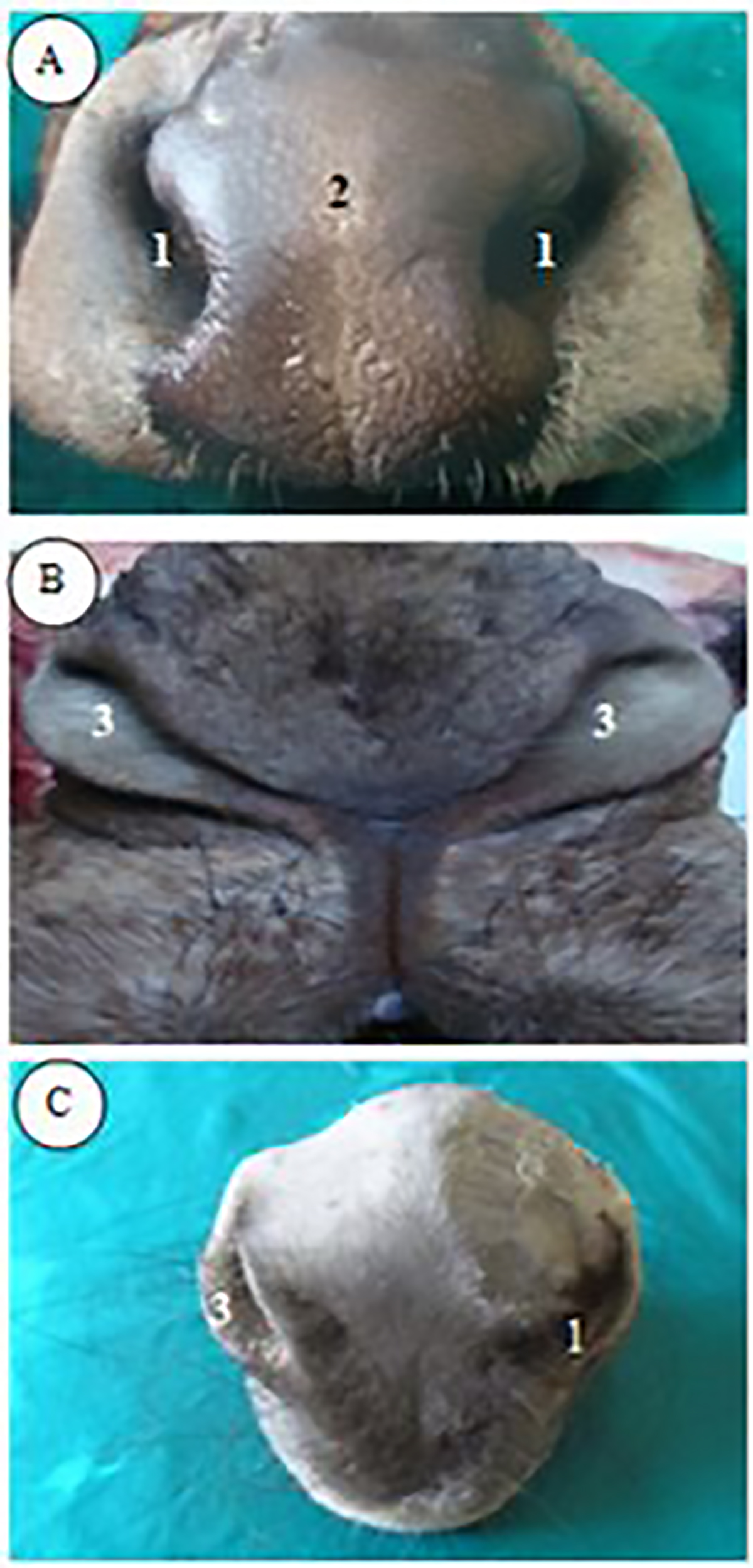
The nostrils, nasal vestibule and vestibular diverticuli The nasal cavity was funnel shaped, opened rostrally by the nostrils (Nares) and caudally by choanae (caudal nares). The nostrils (Fig. 1) was comma shaped in buffaloes, slit like in camels and crescent shaped in donkeys. Moreover, the ventral nasal angle was wider than the dorsal one in buffaloes, while it was narrower than the dorsal one in donkeys and camels. The nostrils of buffaloes were laterally situated while in donkeys they directed obliquely toward the median plane, as the distance between ventral nasal angles was shorter than that of the dorsal nasal angle (Fig. 1). In camels, the longitudinal axis of each nostril was directed rostromedially. These oblique slit-like nostrils in camels formed together with the median labial fissure a characteristic Y- shaped snout (Fig. 1). The nasal cavity divided longitudinally into three parts; the rostral part or the nasal vestibule that covered with hairy skin, the middle part or the proper nasal cavity, which covered with mucous membrane, the later demarcated from the nasal vestibule by limen nasi and lodged by the nasal conchae. The nasal vestibule was extended between nostrils and limen nasi. In camels, it had a lateral pouch named the lateral nasal diverticulum (Fig. 2). It was a paired, cylindrical shaped pouch, extended along the lateral side of the nasal vestibule and located between the rostral parts of the levatorlabii maxillaries, the caninus and the depressor labii maxillaries muscles (maxillolabial group of muscles) and the lateralis nasi muscle. It was also inclined over the nasal process of the incisive bone before piercing the lateral wall of the nasal vestibule. The opening of this lateral diverticulum was located at the level of limen nasi, 1-1.5 cm from the ventral nasal angle and 0.5 cm dorsal to the orifice of nasolacrimal duct. Another vestibular diverticulum was found in donkeys, the false nostrils or the nasal diverticulum. Each one was a funnel shaped pouch supported medially with the lamina of alar cartilage (Fig. 3). Its rostral half was located within the nasoincisive notch, while its caudal half located lateral to the nasal processes of the incisive and maxillary bones. Its lateral wall had an undulating fold, which continued rostrally with the alar fold and divided the pouch partially into rostrodorsal and caudoventral parts. It was related dorsally to the tendon of levator nasolabialis muscle, laterally to the caninus muscle and ventrally to the external nasal branch of the infraorbital nerve.
Fig. 1. A photograph of fresh specimens of rostral end of head in buffalo (A), camel (B), and donkey (C), showing: 1-Nares, 2- Planum nasolabiale, 3- Alae nasi.
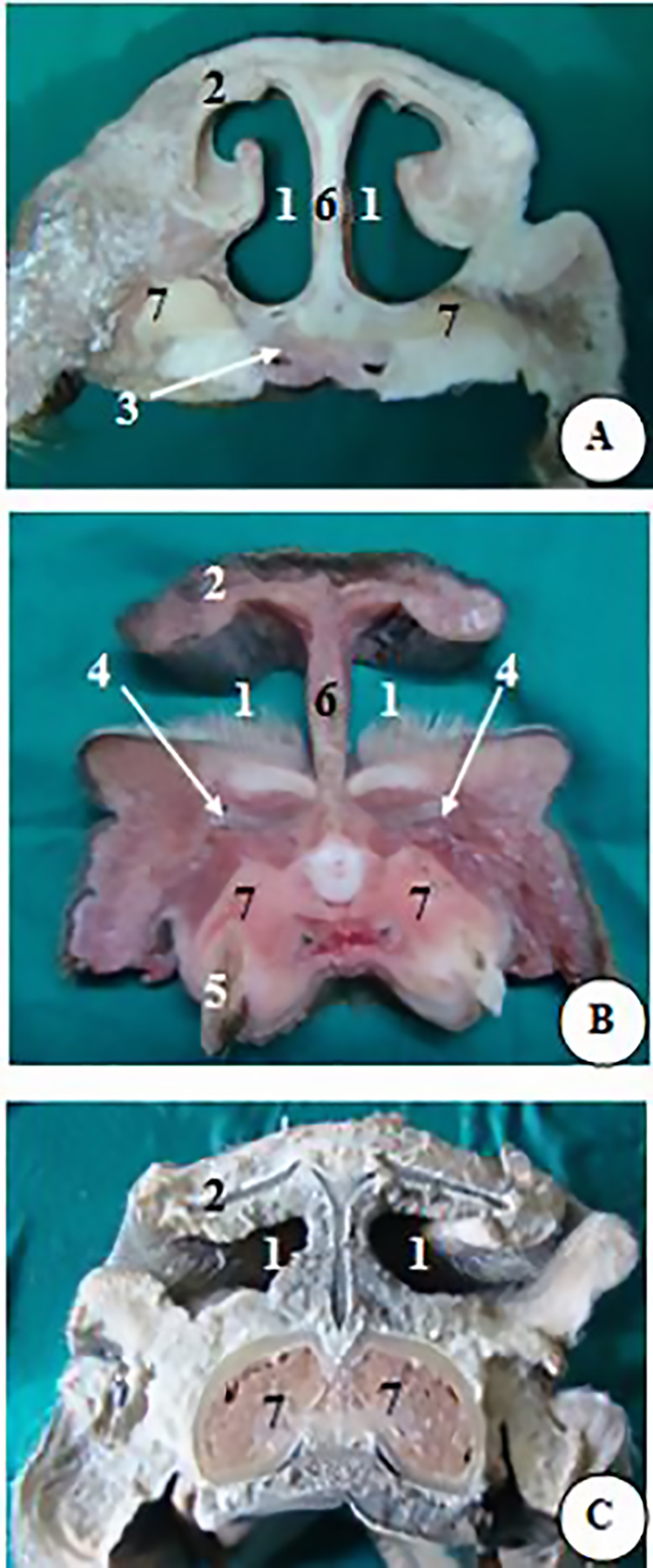
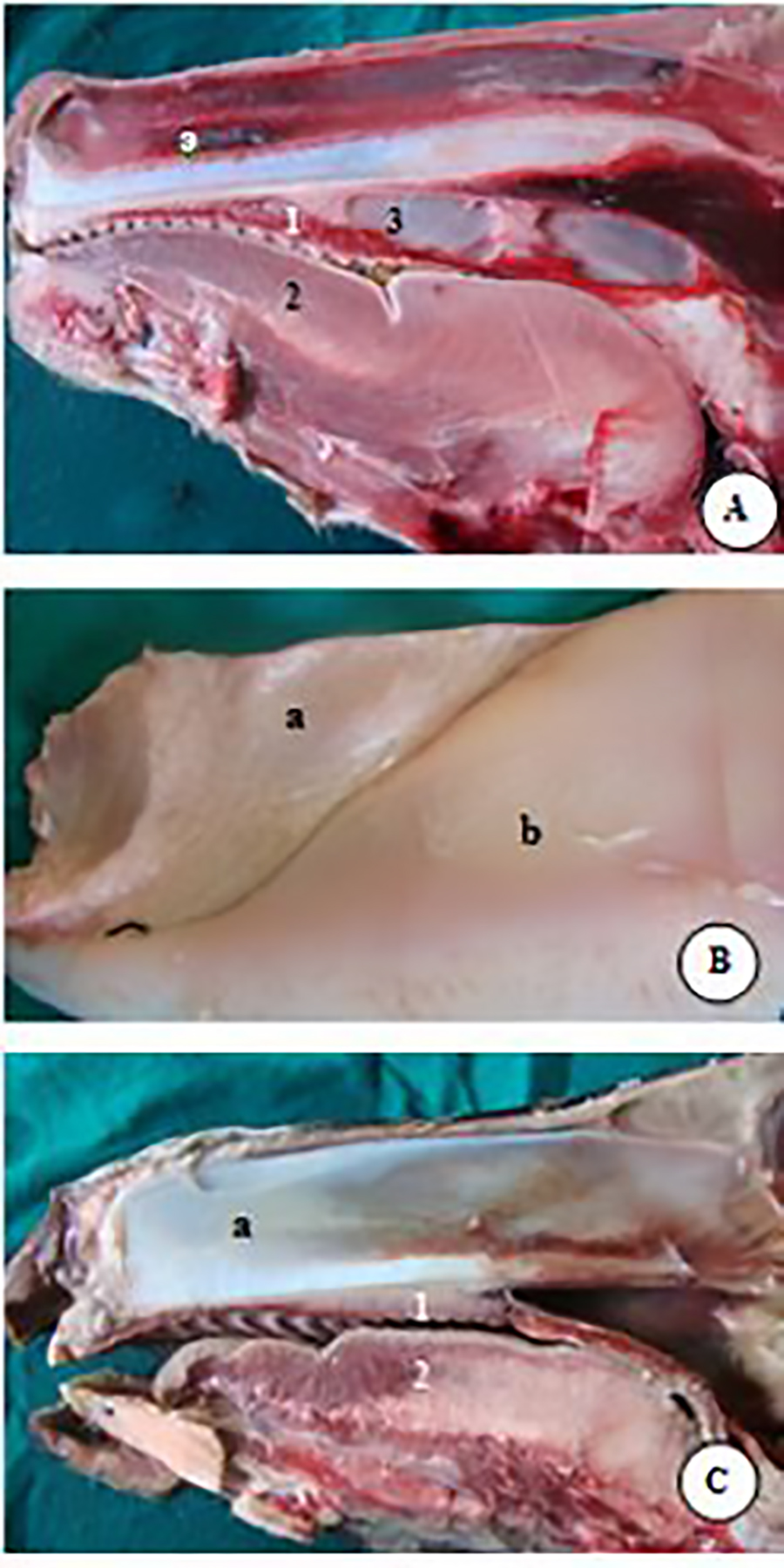
Fig. 2. A photograph of transverse sections of nasal cavity at level of rostral third of dental pad of buffalo (A), incisor III in camel (B) and rostral third of interdental space in donkey (C), showing: 1- Nares, 2- Ala nasi lateralis, 3- Organumvomero nasale, 4- Lateral nasal diverticulum (in camel only), 5- Incisor III, 6- Septum nasi, 7- Os incisivum.
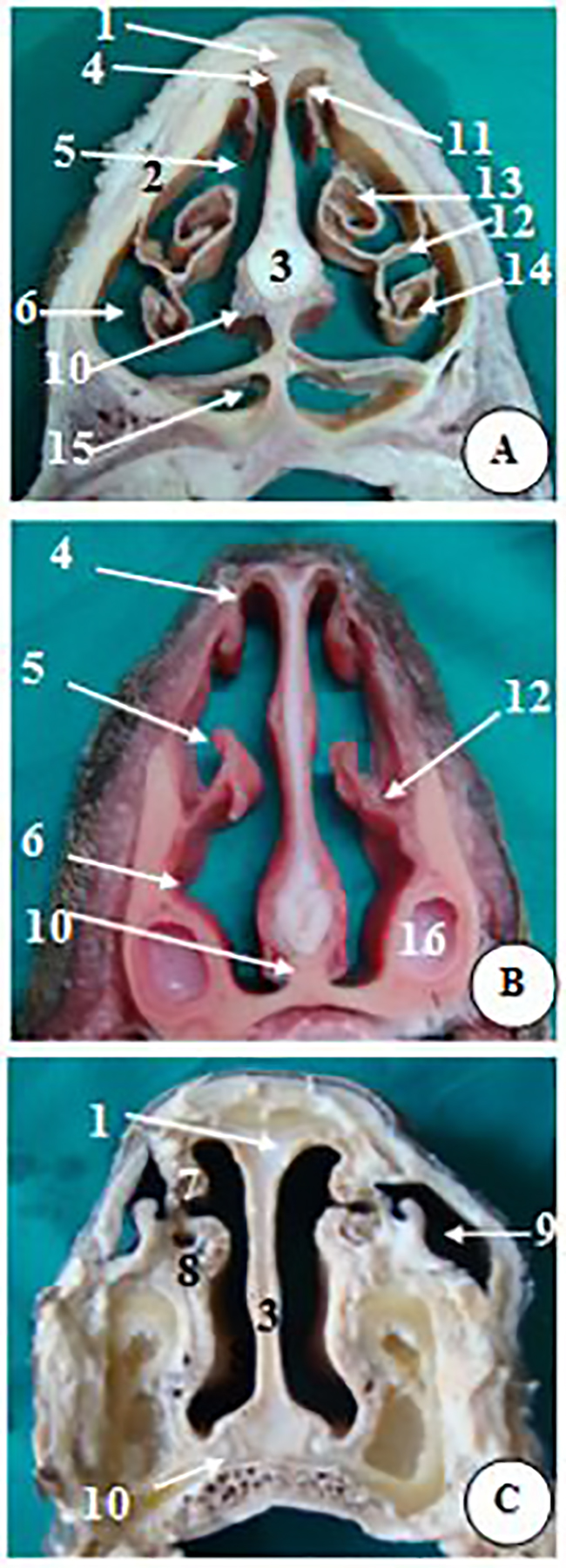
Fig. 3. A photograph of transverse sections of nasal cavity at level of caudal third of interdental space in donkey (C), dental pad of buffalo (A) and first premolar in camel (B), showing: 1- Os nasale, 2- Maxilla, 3- Septum nasi, 4- Meatus nasi dorsalis, 5- Meatus nasi medius, 6- Meatus nasi ventralis, 7- Plica recta, 8- Plica alaris, 9- Diverticulum nasi, 10- Organumvomero nasale, 11- Lamella basalis of dorsal nasal concha, 12- Lamella basalis of ventral nasal concha, 13- Lamella spiralis dorsalis of ventral nasal concha, 14- Lamella spiralis ventralis of ventral nasal concha, 15- Sinus palatines, 16- Root of first premolar tooth in camel. Nasal cartilages (Cartilagines nasi externi) The nasal cartilages were five pairs including, the lateral dorsal nasal, lateral ventral nasal, lateral accessory nasal, medial accessory nasal and alar cartilages (Fig. 4). Camels had the least number of cartilages and the overall size of the cartilaginous skeleton was highly reduced. Only one pair of cartilages was missed in each of buffaloes and donkeys; the alar in the former and the lateral accessory in the later. Whereas, the alar and the ventral lateral nasal cartilages were absent in camels.
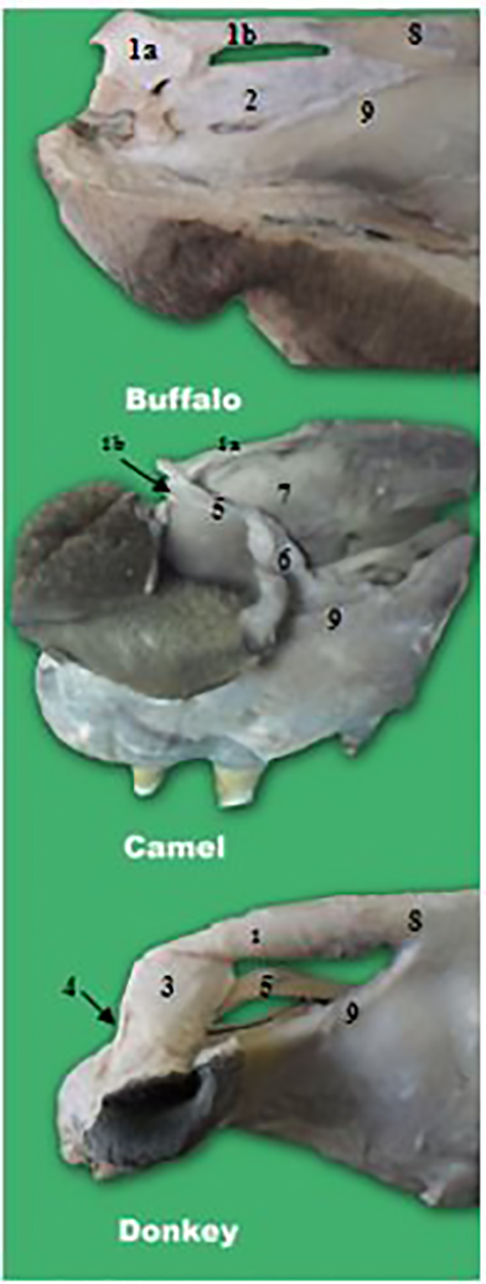
Fig. 4. A photograph of nasal cartilages in buffalo, camel and donkey showing: 1- Cartilago nasi lateralis dorsalis, 1a- Rostral or naric part of Cartilago nasi lateralis dorsalis, 1b- Caudal part of Cartilago nasi lateralis dorsalis, 2- Cartilago nasi lateralis ventralis, 3- Lamina of cartilago alaris 4- Cornu of cartilago alaris, 5- Cartilago nasalis accessoria medialis, 6- Cartilago nasalis accessoria lateralis, 7- Septum nasi, 8- Os nasale, 9- Os incisivum. Dorsal Lateral nasal cartilage (Cartilago nasi lateralis dorsalis) In buffaloes, camels and donkeys, the dorsal lateral nasal cartilage was extended bilaterally along the dorsal border of nasal septum. Additionally, in buffaloes and camels it was attached caudally to the nasal bones, while in donkeys it attached to nasal bones ventrally (Fig. 4). In buffaloes, the dorsal lateral nasal cartilage was 15-17 cm in length and 3.5-4.5 cm in width. In camels, it was 10-11 cm in length and 1-1.5 cm in width, while in donkeys it was 6.5-7 cm in length and 0.6-0.8 cm in width. The dorsal lateral nasal cartilage was undivided in donkeys and had the form of narrow quadrilateral plate of cartilage. In buffaloes and camels, it was divided into rostral and caudal parts (Fig. 4). The rostral or the naric part was irregular quadrilateral and had dorsolateral, rostral, caudal and ventral borders. The rostral and caudal borders were convex dorsally and concave ventrally. The caudal border was attached to ventral lateral nasal cartilage by connective tissue, while the ventral border was attached to the lateral accessory nasal cartilage by connective tissue. In buffaloes, the caudal part of dorsal lateral nasal cartilage was rectangular and directed rostroventrally, while in camels; it was a narrow elongated part that directed caudoventrally. In buffaloes, the dorsal and ventral lateral nasal cartilages were united rostrally and caudally the gap between them was filled by connective tissue rich in blood vessels and fat (Fig. 4). In buffaloes, they formed most of the lateral wall of nasal vestibule, while in camels they were very narrow and the lateral wall of nose was mainly formed of muscles. In donkeys the dorsal lateral nasal cartilage was narrower than that of buffaloes but wider than that of camels and the lateral wall of nose was formed mainly of skin. Ventral Lateral nasal cartilage (Cartilago nasi lateralis ventralis) In buffaloes, the ventral lateral nasal cartilage was well developed while in donkeys it was ill developed but in camels, it was absent (Fig. 4). In buffaloes, it was in the form of quadrilateral plate attached to the dorsal part of the nasal process of incisive bone with its caudal part wedged in the nasoincisive notch. In donkeys, it was extended from the ventral border of the cartilaginous part of nasal septum and filled the palatine fissure. In buffaloes, the ventral border was curved medially and attached to the medial accessory nasal cartilage. In buffaloes, the ventral lateral nasal cartilage was 10-12 cm in length and 3 - 4 cm in width, while in donkeys it was 2.5 -3 cm in length and 0.3-0.5 cm in width. Lateral accessory nasal cartilage (Cartilago nasalis accessoria lateralis) In camels and buffaloes, it was anchor shaped cartilage with a lateral lamina that supported the lateral nasal wing. It also had a medial curved part that attached to the naric part of the dorsal lateral nasal cartilage (Fig. 4). In buffaloes, the lateral accessory nasal cartilage was 2.5 - 3 cm in length, while in camels; it was 1.5-2 cm. This cartilage was absent in donkeys. Medial accessory nasal cartilage (Cartilago nasalis accessoria medialis) In buffaloes, camels and donkeys, it was a small cartilage lodged within the alar fold. In buffaloes, it was raised from the ventral border of the ventral lateral nasal cartilage, while in camels it was attached to the dorsal lateral nasal cartilage and lateral accessory nasal cartilage (Fig. 4). In donkeys, it was attached to the basal fold of the ventral nasal concha. In buffaloes, it was an irregular quadrilateral cartilage with a convex medial surface, but in camels and donkeys it was S shape. In buffaloes, the medial accessory nasal cartilage was 2-3 cm in length. In camels, it was small of 2-2.5 cm in length, while in donkeys it was larger comparing to the size of the head (3-4 cm). Alar cartilage (Cartilago alaris) The alar cartilage was present only in donkeys. It was a comma shaped cartilage that formed of two parts, the dorsal part or the lamina and the ventrolateral part or the cornu (Fig. 4). The lamina was supported the medial wing of the nostril and the rostral part of alar fold. It was attached caudally to the dorsal lateral nasal cartilage. The cornu was in the form of curved rod that supported the ventral nasal commissure. The lamina was 2.5-3 cm in length, while the cornu was 1.5-2 cm in length. The alar cartilage and its fellow were located back to back and attached by connective tissue. The rostral border of lamina of alar cartilage articulated dorsally with rostral end of cartilaginous part of nasal septum by a synovial joint. Nasal septum (Septum nasi) In buffaloes and donkeys, the nasal septum was formed of a rostral cartilaginous part and a caudal osseous part. However, in camels, it was formed of three parts, the rostral mucomuscular, the middle cartilaginous and the caudal osseous parts (Fig. 5). In buffaloes, the mucous membrane covering nasal septum has dorsal and ventral septal swelling bodies. The dorsal swelling body was ridge or crest like structure which located along middle of the lateral surface of the nasal septum opposite to the middle nasal meatus. The ventral septal swelling body was located on either side of the ventral border of the nasal septum. It was more developed than the dorsal one and decreased in size caudally.
Fig. 5. A photograph of fresh specimens of nasal septum in buffalo (A), camel (B), and donkey (C) showing: a- Cartilago septi nasi, b- Muscular part of nasal septum. 1- Palatum durum, 2-Lingua,3- Sinus palatinus. Vomeronasal organ (Organumvomero nasale) The vomeronasal organ was a tubular structure covered by mucous membrane of the nasal cavity on both sides of the nasal septum (Fig. 6). Its beginning was located caudal to the ventral nasal angle by 3 cm in buffaloes, 4-5 cm in camels and 5-6 cm in donkeys. Its caudal blind end was located caudal to the ventral nasal angle by 13-14 cm in buffaloes, 20-21 cm in camels and 13-14 cm in donkeys. The blind end was also at the level of the second premolar tooth in donkeys, the third premolar tooth in buffaloes and the fourth premolar tooth in camels. The length of the vomeronasal organ was 10- 11 cm in buffaloes, 16-17 cm in camels and 8-9 cm in donkeys. Its diameter increased gradually toward the caudal part and was about 0.5-1cm in buffaloes 0.4- 0.5 cm in camels and 0.3-0.4 cm in donkeys. Nasal conchae (Conchae nasalis) The nasal conchae in buffaloes, camels and donkeys included the dorsal nasal concha, the middle nasal concha, the ventral nasal concha and the ethmoidal conchae. In buffaloes and donkeys, the nasal concha was extended through the whole length of the nasal cavity (Fig. 6). However, in camels they did not extend to the rostral third and condensed only in the caudal two thirds. Only the straight alar and basal folds were found rostrally (Fig. 6). In buffaloes and camels, the ventral nasal concha divided into pars dorsalis and pars ventralis, these parts in camels were S shaped (Fig. 7). In donkeys, the dorsal and the ventral nasal conchae divided into pars rostralis and pars caudalis by septum conchae dorsalis and septum conchae ventralis. These septa located at the level of third premolar tooth (Fig. 7). Dorsal nasal concha (Concha nasalis dorsalis) In buffaloes and camels, the dorsal nasal concha was wide in the middle and tapered toward either ends. In donkeys, it was funnel shaped, wide caudally and narrowed rostrally. In buffaloes and donkeys, it was the longest concha, while in camels it was shorter and smaller than the ventral nasal concha. Its length was 11-12 cm in buffaloes, 13-14 cm in camels and 10-11cm in donkeys. In buffaloes its height was measured 2-3 cm at the middle widest part and 0.3-0.4 cm at the narrowest part (its end). In camels, its height was 3-4 cm at the middle widest part and 1-2 cm at the narrowest parts. In donkeys, the height was 3-4 cm at the middle widest part and 0.7-1 cm rostrally near straight fold. In buffaloes and camels, the rostral two thirds of nasal concha were formed of a basal lamella (Figs 2, 3). While, the caudal third enclosed the dorsal conchal sinus. In donkeys, the rostral third enclosed a recess, the middle third contained recess and bulla, and the caudal third enclosed the dorsal conchal sinus.
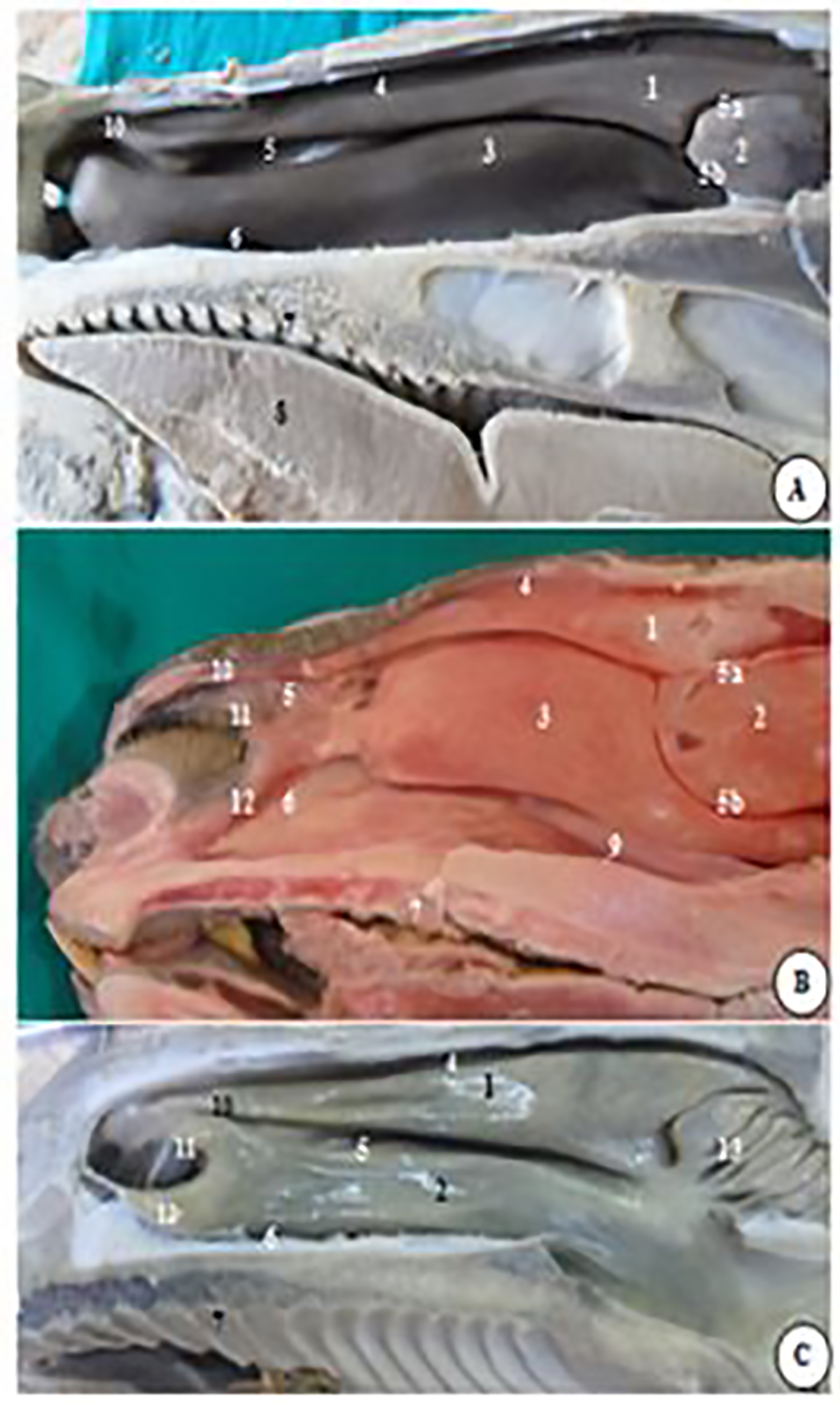
Fig. 6. A photograph of formalized specimens (A, C) and fresh specimen(B) of right paramedian section of nasal cavity in buffalo (A), camel (B) and donkey(C) showing: 1- dorsal nasal concha, 2- middle nasal concha, 3- ventral nasal concha, 4- dorsal nasal meatus, 5- middle nasal meatus, 5a- dorsal branch, 5b- ventral branch, 6- ventral nasal meatus, 7- hard palate, 8- tongue, 9- vomeronasal organ, 10- straight fold, 11- alar fold, 12- basal fols, 13- Ethmoidal conchae.
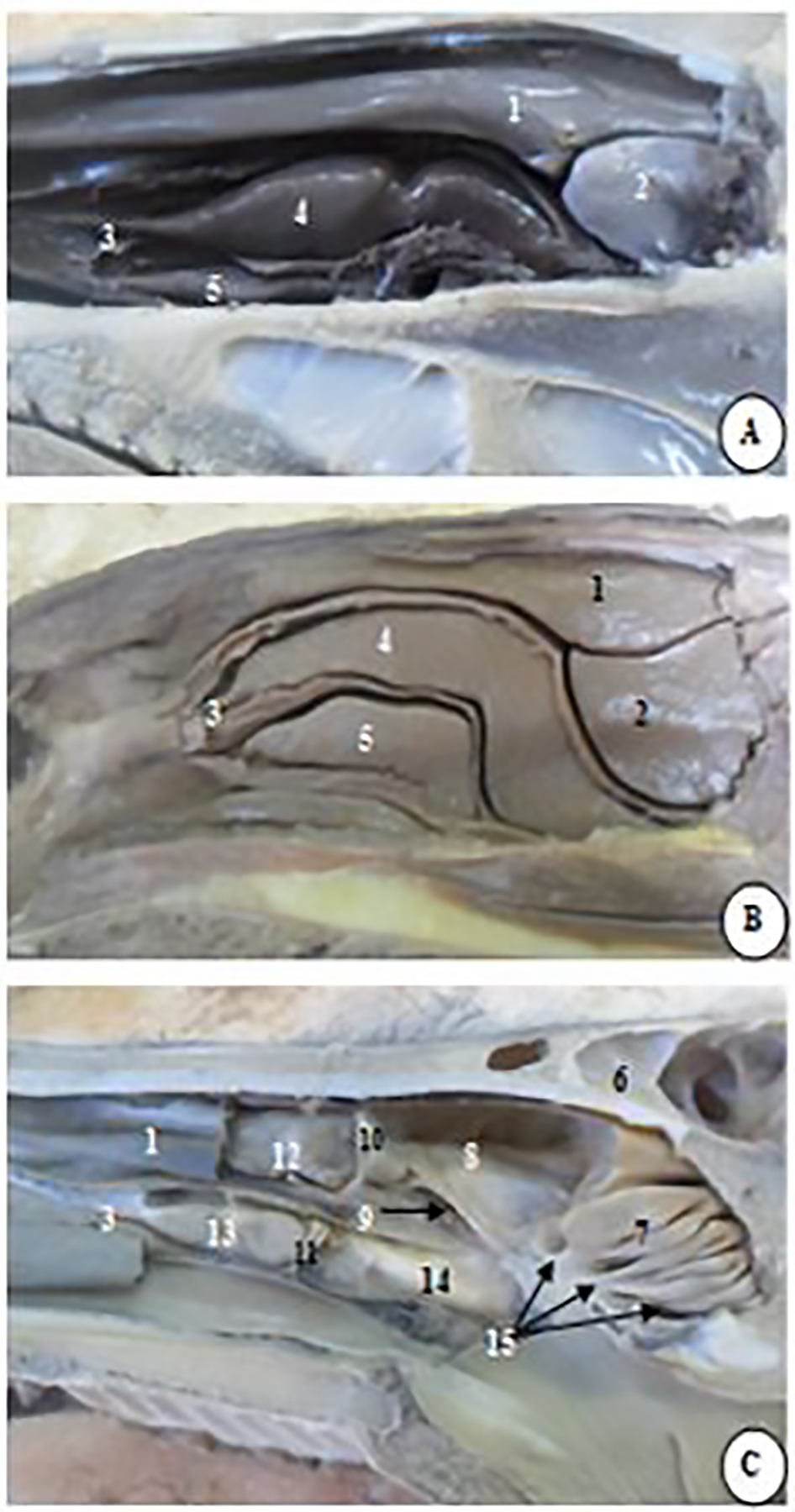
Fig. 7. A photograph of formalized specimens of right paramedian section of nasal cavity in buffalo (A), camel (B) and donkey(C) showing: 1- Concha nasalis dorsalis (opened in donkey), 2- Concha nasalis media, 3- Concha nasalis ventralis (opened), 4- Pars dorsalis of ventral nasal concha, 5- Pars ventralis of ventral nasal concha, 6-Sinus frontalis, 7- Conchae ethmoidal, 8- Sinus concho frontalis, 9- Apretura naso maxillaris, 10- Septum conchae dorsalis, 11- Septum conchae ventralis, 12- Bulla conchalis dorsalis, 13- Bulla conchalis ventralis, 14- Sinus conchalis ventralis, 15- Meatus ethmoidales. Straight fold (Plica recta) The straight fold was the rostral continuation of dorsal nasal concha along lateral wall of the nasal cavity toward nostril. In donkeys, the beginning of the straight was appeared as two rounded ridges, which supported by cartilages. Continuing rostrally, these two ridges were rejoined again to continue as one straight fold (Figs. 6, 7). In buffaloes, it was also contained a cartilaginous prolongation of the dorsal nasal concha. While in camels, no cartilaginous prolongation was found. Its length was 4-5 cm in buffaloes, 7-10 cm in camels and 9-10 cm in donkeys. Its diameter was 0.5-1 cm in buffaloes, 0.5- 1 cm in camels and 0.5-1.5 cm in donkeys. Ventral nasal concha (Concha nasalis ventralis) The ventral nasal concha had two rostral folds, the alar and the basal folds. In camels, these folds did not arise from the ventral nasal concha directly. The concha was extended for about 1.5-2 cm as a single constricted part (1.5-2 cm), which bifurcated rostrally to form the alar and the basal folds (Figs. 6, 7). In buffaloes and donkeys, the ventral nasal concha was shorter and broader than the dorsal nasal concha. While in camels, it was the longest and the largest concha. Its length was 25-28 cm in buffaloes, 15-20 cm in camels and 17-19 cm in donkeys. In buffaloes, the rostral and the middle parts were nearly equal in height and was 3-4 cm, with the widest part located caudally (6-7 cm). In buffaloes and camels, the rostral third of the ventral nasal concha was formed of a basal lamella which carried dorsal and ventral spiral lamellae (Figs. 2, 3). In the middle third, these spiral lamellae were enclosed recess and bulla, while the caudal third of that concha enclosed the ventral nasal conchal sinus. In camels, the caudal third of the ventral nasal concha was twisted and the ventral spiral lamella was situated lateral to the dorsal one. In donkeys, the rostral third enclosed a recess, the middle third was contained recess and bulla, whereas, the caudal third enclosed the ventral conchal sinus. Alar fold (Plica alaris) In buffaloes and camels, the alar fold was extended along the lateral wall of the nasal vestibule parallel to rostral part of dorsal nasal concha and the straight fold, while in donkeys, it extended from the concha to the medial wing of nostril (Figs. 6, 7). In the three species, the alar fold was supported by the medial accessory cartilage. In buffaloes, the alar fold was lodged in the terminal part of the nasolacrimal duct. Basal fold (Plica basalis) In the three species, the basal fold was rounded in cross section and presented thick submucous venous plexuses. Its length was 1-2 cm in buffaloes, 4-5 cm in camels and 2-3 cm in donkeys. Its diameter was 0.5-1 cm in buffaloes, 0.3- 0.5 cm in camels and 0.5-1.5 cm in donkeys. In camels and donkeys, it contained the terminal part of nasolacrimal duct. Middle nasal concha (Concha nasalis media) In buffaloes and camels, the middle nasal concha was formed by endoturbinate II (4/2). It was lodged between the caudal parts of the dorsal and ventral nasal conchae and enclosed the middle conchal sinus (18/9, 19/5). However, in donkeys, it was not recognized because of the nearly equal lengths of ethmoidal conchae (4/13). Its length was 7-9 cm in buffaloes and 9-10 cm in camels. Its height was 3.5-4 cm in buffaloes and 4.5-5 cm in camels. Ethmoidal conchae (Conchae ethmoidales) The ethmoidal conchae were occupied the caudodorsal part of nasal fundus (4/13, 6/7, 7/2, 8/5). The ethmoidal conchae were divided into endoturbinates and ectoturbinates. The latter were smaller and arranged lateral to the endoturbinates. In buffaloes and donkeys, the endoturbinates were arranged in one row dorsoventrally. In camels, they were arranged in three rows mediolaterally as follows, the medial row (3 conchae); the intermediate (4 conchae) and the lateral, which was the largest and has one concha. The ectoturbinates was 11-15 in buffaloes, 6-7 in camels and 8-10 in donkeys. Nasal meatuses The nasal conchae divided the nasal cavity into dorsal, middle and ventral nasal meatuses (4/4 and 5&6). In buffaloes and camels, the middle nasal concha was divided the middle nasal meatus caudally into dorsal and ventral branches (4/5a and 5b). The dorsal nasal meatus was bounded dorsally by nasal bone and ventrally by dorsal nasal concha and terminated at junction of frontal bone and cribriform plate of ethmoid bone. Its length was 27-30 cm in buffaloes, 23-26 cm in camels and 20-23 cm in donkeys. The middle nasal meatus was wider than the dorsal nasal meatus and narrower than the ventral nasal meatus and led directly into choanae. It contained the openings of vomeronasal organ and incisive duct and the nasomaxillary opening. The common nasal meatus was extended between the nasal septum medially and the nasal conchae laterally and extended between the roof and floor of the nasal cavity (13/16, 14/18, 15/10, 16/7, 17/6). There were also three principle ethmoidal meatuses between endoturbinates of ethmoidal conchae (6/15). Choanae The nasal cavity was communicated caudally with the nasopharynx through the choanae or caudal nares (19/18, 20/8). In buffaloes and donkeys, it was oval, while in camels it was nearly circular in outline. In buffaloes and camels, it was single, but in donkeys it was divided into two equal parts by vomer bone. In camels and donkeys, it was nearly horizontal to the vertical plane, while in buffaloes it was slightly oblique. |
|
|
|
Discussion |
|
The results of this study revealed the anatomical peculiarities in each species, which can be summarized as follows; in buffalo and donkey, the arrangement of the nasal choncae was similar to that described in other domestic animals (Hare, 1975; Nickel et al., 1979; Walker, 1982; Abdel–Aziz, 1983; Evans and Delahunta, 2000). However, in camels, the choncal arrangement and the twisting of the ventral nasal concha were described only in proboscis-bearing mammals, such as Saiga antelope (Saiga tatarica) and Mooze (Alces alces) (Clifford, 2003). Previous study on dromedary camels had been confirmed this conchal arrangement (Gewaily, 2009). Recent studies have been also confirmed the anatomical relations between camels and the group of mammals known as “proboscis-bearing mammals” (Eshra and Badawy, 2014). As both of them have a muscular group that may enable the nostrils to close in adverse climatic conditions (Clifford, 2003; Eshra and Badawy, 2014). Camels also have a nasal plane similar to this group of animals (Eshrah, 2017). For these, the authors suggested that the nasal anatomy in camels may be related to the proboscis bearing mammals. Supporting this idea is that they sharing several anatomical features, which including, the presence of muscular conformation of the nasal aperture (Eshra and Badawy, 2014), the reduction of the bony and cartilaginous skeleton (Badawi and Fateh El-Bab, 1974; lateral Gewaily, 2009), the presence of vestibular recesses known as the lateral nasal diverticulum (Smuts and Bezuidenhout, 1987). The later is similar to the vestibular recesses found in Saiga antelope (Clifford, 2003). However, further investigations are still required to test this hypothesis. Results also revealed that buffaloes have a set of nasal cartilage that was completely surrounded the narial aperture. However, this greatly differs from that found in donkeys and camels, as they have a reduced cartilaginous skeleton. In donkeys, the lateral wall of the nasal cavity was nearly devoid of cartilages. Instead of having a lateral cartilaginous wall, donkeys have a vestibular diverticulum known as nasal diverticulum (Hare, 1975; Hamoda, 2000). This may give more space for the lateral nasal wall to expand during forced inspiration, helping the animal to increase the respiratory rate. In camels, the reduction of nasal cartilages was substituted by extensive muscular attachment (Eshra and Badawy, 2014). Another peculiarity in camel narial anatomy was that the rostral third of nasal septum was formed entirely of muscles and covered with mucus membrane (Badawi and Fateh El-Bab, 1974; Gewaily, 2009). As mentioned above, this may be related to special mechanism for narial closure similar to that found in proboscis bearing-mammals. In the three animals, the mucous membrane covering the nasal septum has dorsal and ventral septal swelling bodies. The vomeronasal organ was also located on both sides of the nasal septum. It was notably longer and wider in camels (Badawi and Fateh El-Bab, 1974). The blind end of the vomeronasal organ was located at the level of the second premolar in donkeys, this similar to the finding of Hamoda (2000) and Mansour et al. (2001). In buffaloes, it was at the level of the third premolar as observed by Fouad et al (1984) in the same animals and by Besoluk et al. (2001) in Angora goat. In camels, it was located at the level of the fourth premolar this finding was similar to that observed by Gewaily (2009). Based on the results of this study, the dorsal nasal concha was the longest concha in buffaloes and donkeys, this agree with that mentioned by Nickel et al. (1979) in ruminants and equine. Conversely, in camels, it was shorter and smaller than the ventral nasal concha. Similar findings were described by Badawi and Fateh El-Bab (1974) and Gewaily (2009) in the same animal. It is important to put in consideration that the length of mucosal folds was excluded in this study and the conchal length was the extension of its turbinate bone. In the examined three species, the straight fold was also of different extensions and forms. In buffaloes and camels, it was straight as its name implied. In donkeys, it was in the form of two rounded ridges, supported by cartilages and rejoined again to continue as one straight fold, these results were similar to that reported by Hare (1975) in horse and Hamoda (2000) in donkeys. In buffaloes, it also contained a cartilaginous prolongation, while in camels no cartilage was found. In cross sectional anatomy, the dorsal nasal concha was simple as any other endoturbante from the ethmoidal system of conchae. In contrast, the ventral nasal concha was complex and has different shape according to species (Schaller, 2007). Although in buffaloes and camels, the ventral nasal concha was divided into dorsal and ventral parts, these parts in camels were S shaped. This twisting allows condensation of the nasal conchae in the caudal two thirds and explains why this concha was shorter in camels. The plane of the choanae, the caudal opening of the nasal cavitywas nearly horizontal in camels (Hare, 1975; Gewaily, 2009), and in donkeys (Hamoda, 2000), while that of buffaloes was slightly oblique (Abdel-Aziz, 1983). The choanae was also divided into two equal parts by vomer bone in donkeys while in buffaloes and camels it was single. |
|
|
|
Conclusion |
|
In contrast to buffaloes and donkeys, which follow the anatomical plan of their ruminant and equine tribes, camel nasal cavity presented interesting anatomical features similar to those of proboscis-bearing mammals and differed with those of other domestic animals. |
|
|
|
Conflict of Interests |
|
|
|
Authors declared that no conflict of interests exist. |
|
|
|
References |
|
|
|
Abdel-Aziz, S.E., 1983. Some anatomical studies on nasal cavity of buffalo (Bos bubalis) in Egypt. M.V.SC. Thesis. Zagazig Fac. of Vet. Med. Zagazig University, Egypt. Ahmed, A.A.M., Moussa, E.A.M., Osman, A.H.K., 2005. Anatomical and histological studies on the nasal cavity of the dromedary camel (Camelus dromedarius). 4th Int. Sci. Conf., Mansoura University. pp. 815-840. Badawi, H., Fateh El-Bab, M.R., 1974. Anatomical and histological studies on the nasal cavity of the camel (Camelus dromedarius). Assiut Vet. Med. J. 1, 1-14. Besoluk, K., Eken E., Boydak, M., 2001. The vomeronasal organ in Angora goat caprahircus. Vet. Arhiv. 71, 11-18. Clifford, A.B., 2003. Narial novelty in mammals: Case studies and rules of construction. M.Sc. Faculty of the college of arts and science, Ohio University, U.S.A. Dyce, K.M., Sack, W.O., Wensing, C.J.G., 1996. Textbook of veterinary.2nd ed. W.b. Saunders Company, Philadelphia, London, Toronto, Montreal, Sydney, Tokyo. Eshra, E.A., Badawy, A.M., 2014. Peculiarities of the camel and sheep narial musculature in relation to the clinical value and the mechanism of narial closure. Indian Journal of Veterinary Anatomy 26, 10-13. Eshrah, A.E., 2017. The Camel Rhinarium: A study revealing the presence of the nasal plane in dromedary camel (Camelus dromedarius), with special reference to its epidermal structure. Anat. Histol. Embryol. 46, 65-72. Evans, H.E., Delahunta, A., 2000. Guide to the dissection of the dog .9th ed. W.B. Saunders Company. Philadelphia. U.S.A., pp. 275-290. Fath El-Bab, M.R.M., 1970. Anatomy and histology of the respiratory system of the camel. M.D. Thesis, Assiut Fac. Vet. Med., Assiut University, Egypt. Fouad, S.M., Ewais, M.S., Abdel-Aziz, S.E., Mobarak, A.N., 1984. Vomero-organ of buffaloes in Egypt Bos bubalis. Vet. Med. J, 32, 147-160. Gewaily M.S.M., 2009. Some anatomical studies on the nasal cavity in the one humped camel (Camelus dromedarius). M.V.Sc. Thesis, Faculty of Vet. Med. Kafrel-sheikh Univ. Egypt. Hamoda, H.S.A., 2000. Some anatomical studies on the nasal cavity of the donkey. M.V.SC. Thesis, Kafr El-Sheikh Fac. Vet. Med., Tanta University, Egypt. Hare, W.C.D., 1975. Equine respiratory system and ruminant respiratory system. In Sisson and Grossman. Anatomy of the domestic animals 5th ed. W.B. Sounders Company, Philadelphia. U.S.A. 1, 498-524, 916-937. Mansour, A.A., Ali, M.A., Hamoda, H.S.A., 2001. Some morphological studies on the vomeronasal organs of the donkey. Assiut Vet. Med. J. 45, 14-23. Nickel, R., Shummer, A., Seiferle, E., 1979. The viscera of the domestic animals 2nd revised ed. Verlag Paul Parey. Berlin, Hamburg. pp. 211-281. Nomina Anatomica Veterinaria, 2006. Electronic edition. Published by the international committees on veterinary gross anatomical nomenclature under the financial responsibility of the world association of veterinary anatomists. Zurich and Ithaca, New York. Schaller, O., 1992. Illustrated Veterinary Anatomical Nomenclature. 5th ed. Ferdinand Enke. Published by i. b. d., Limited. Smuts, M.S., Bezuidenhout, A.J., 1987. Anatomy of the dromedary. 1st ed. Clarendon Press, Oxford. U.S.A. PP.105-121. Thiemann, A.K., Bell, N.J., 2001. The peculiarities of donkey respiratory disease. International Veterinary Information Service, Ithaca, New York, U.S.A. Verlag Stuttgart, Germany, pp. 174-194. Walker, W.F., 1982. A study of the cat, with references to human beings. 4th ed. Sounders College Publishing, U.S.A., pp. 152-159. |
|
|