|
|
Journal of Advanced Veterinary Research Volume 9, Issue 3, 2019, Pages: 130-132 www.advetresearch.com |
|


|
Management of Ocular Setariasis in Ponies with Local Ocular Ivermectin Injection |
|
|
|
Weerasekara M.N.K. Jayathilake, Kalaichelvan Nizanantha, Luwisdura N.A. De Silva |
|
Department of Farm Animal Production and Health, Faculty of Veterinary Medicine and Animal Science, University of Peradeniya, Peradeniya, Sri Lanka |
|
Received: 27 April 2019; Accepted: 11 July 2019 |
|
*Corresponding author: |
|
Abstract |
|
Equine ocular Setariasis is a vision threatening disease of equine, caused by Setaria spp. This clinical communication describes two equine ocular Setariasis cases which were successfully treated with a single dose of ocular Ivermectin injection. Two ponies were referred to the Ambulatory clinic, Faculty of Veterinary Medicine, Peradeniya, Sri Lanka with the complaint of observation of a moving worm inside the anterior chamber of the eye since few days. The cloudiness of the cornea has been gradually increasing since the day the owner observed the worm inside the eye. Upon the clinical examination, both ponies were normal except the signs related to the eye condition including extent lacrimation, and moderate, unilateral corneal opacity. On close examination of the affected eye, the swirling movements of white thread like worm swimming continuously in aqueous humor of anterior chamber of the eye was evident in both cases. The ponies were properly restrained. 0.05ml of 1% ivermectin was loaded in to a sterile 1cc syringe with a 29G needle and was double diluted with normal saline. While the pony’s head was held firm, the needle was inserted in to the anterior chamber of the eye and the drug was released slowly. Prophylactic eye ointment containing an antibiotic i.e. 3% Tetracycline Hydrochloride (Galentic®) ointment was prescribed to be applied in the eye twice daily. Within an hour after the ivermectin treatment, the movements of the worm became sluggish and the movements were restricted to the most ventral part of the anterior chamber. The worm which was in the anterior chamber died on the same day of treatment. But few days later, the dead worms had triggered an inflammatory reaction in the eye, thus the animals were treated with an ophthalmic preparation containing NSAID i.e. 0.5% Ketorolac Tromethamine (Ketrosan®) ophthalmic solution. The condition recovered without any impairments of the vision or damages to the eye. Treatment of ocular Setariasis is always being a challenge. There are reported cases of successful surgical and medical interventions for ocular Setariasis in equids. This method would provide a less invasive, quick method done using only a single dose of ocular ivermectin injection in order to treat equine ocular Setariasis. |
|
|
|
Keywords: Equine, Ivermectin, Ocular Setariasis, Setaria Digitata |
|
|
|
Background |
|
|
|
Equine ocular Setariasis is a vision threatening disease of equine, caused by Setaria spp, a genus of filarial worms (Gangwar et al., 2008). S. digitata is a parasite of cattle and hoofed animals and is found mainly in Asia. S. equina infects horses and other equids worldwide. Even though the predilection site of adult Setaria worms is the peritoneal cavity, the parasite exhibits migratory behavior into various organs such as heart, lung, spleen, kidney, uterus, oviduct, ovary, urinary bladder and occasionally they can get into the central nervous system or the eyes (Pratap et al., 2005; Yadav et al., 2006). Adult female worms release microfilariae in the abdominal cavity of their hosts. Mosquitoes become infected with microfilariae when they feed blood of infected hosts and the microfilariae develop to infective larvae inside the mosquitoes in 2 to 3 weeks (Rafee and Amarpal, 2016) . The infected mosquitoes then transmit these infective larvae to other susceptible hosts during their blood meals (Rafee and Amarpal, 2016). The prevalence of ocular Setariasis is high when the mosquito vector population is high. All equines are generally more prone for ocular worm (Pratap et al., 2005). The serrated cuticle of the worm, erratic movements and toxins liberated by the dead worms within the anterior chamber of the eye causes severe irritation and trauma followed by inflammation in the cornea leading to corneal opacity, corneal edema, cataract and retinal detachment (Gopinathan et al., 2013; Rafee and Amarpal, 2016). The dead filarial worm may get attached to the endothelium in the anterior chamber eventually resulting in blindness (Jaiswal et al, 2006). The affected animals might display signs of lacrimation, photophobia, corneal opacity, conjunctivitis and even loss of vision in cases when treatment is delayed (Patil et al., 2012; Gopinathan et al., 2013) . It is commonly unilateral, but bilateral occurrence is also reported (Patil et al., 2016). Diagnosis of the condition can be done by the clinical signs and close inspection of the eye. Hematological tests such as full blood count, microscopic examination of wet blood films and Knott’s test can also be used as supplementary diagnostic tests (Rafee and Amarpal, 2016). Both medical and surgical treatment have been successful for the condition (Tuntivanich and Tiawsirisup, 2010). |
|
|
|
Case Presentation |
|
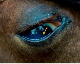
Two ponies were referred to the Ambulatory clinic, Faculty of Veterinary Medicine, Peradeniya, Sri Lanka with the complaint of observation of a moving worm inside the anterior chamber of the eye since few days. The history includes an increased cloudiness of the cornea since the day they observed the worm inside the eye. Complete general clinical examination was performed for both cases including a thorough ophthalmic examination. Other parameters of both ponies were normal except the signs related to the eye condition including extent lacrimation, and unilateral corneal opacity. The corneal opacity in both cases caused by the worm was graded as moderate, a cloudiness of the cornea was observed with visible worm. The anterior chamber was thoroughly examined to detect the presence of worm/s inside the chamber. On close examination of the affected eye, the swirling movement of white thread like worm swimming continuously in aqueous humor of anterior chamber of the eye was evident in both cases (Figure 01). Therefore, the condition was diagnosed as ocular Setariasis due to the possibility of detecting Setaria worms inside the anterior chamber of the aberrant host. The animals were handled in accordance with the institutional guidelines and ethics for care and use of animals at the University of Peradeniya. |
|
Fig. 1. Setaria worm in the anterior chamber of the eye (Yellow Arrow) |
|
Treatment and Management |
|
The ponies were properly restrained. 0.05ml of 1% ivermectin (IVOMEC®, Merial Saude Animal Ltda., Brazil) was loaded in to a sterile 1cc syringe with a 29G needle and was double diluted with normal saline. While the pony’s head was held firm, the needle was inserted in to the anterior chamber of the eye and the drug was released slowly. Gently the needle was withdrawn back. The ponies were closely observed for several hours after the treatment for any hypersensitive reactions. Prophylactic eye ointment containing an antibiotic i.e. Tetracycline Hydrochloride ointment USP 3% (Galentic Pharma Pvt. Ltd., India) was prescribed to be applied in the eye twice daily. |
|
|
|
Prognosis Within an hour after the treatment, the movements of the worm became sluggish and the movements were restricted to the most ventral part of the anterior chamber. The prognosis of both of the cases were good and the follow up of the cases revealed that the animals have recovered the condition. The worm which was in the anterior chamber had died on the same day of treatment. But the dead worms had triggered an inflammatory reaction in the eye, thus the animals were treated with an ophthalmic preparation containing NSAID i.e. Ketorolac Tromethamine 0.5% ophthalmic solution (Ketrosan®, Sante Pvt. Ltd., Pakistan). The condition recovered without any impairments of the vision or damages to the eye. |
|
|
|
Discussion |
|
Equine ocular Setariasis is a vision threatening disease of equine, caused by Setaria spp, a genus of filarial worms (Gangwar et al., 2008). Occasionally the parasite exhibits migratory behavior into the eyes causing ocular Setariasis (Pratap et al., 2005; Yadav et al., 2006). Definitive diagnosis of the condition can be done by a complete ophthalmic examination, which reveals moving worm in the anterior chamber of the eye. If worms are not visible in the anterior chamber microscopic examination of wet blood films and Knott’s test can be performed to detect the microfilariae of the Setaria species (Rafee and Amarpal, 2016). Furthermore, anemia with leukocytosis in complete blood count, and an accelerated erythrocyte sedimentation rate (ESR) has been reported in previous studies and can be used as supplementary tests for the diagnosis of the condition (Muhammad, 2007). In both of our cases, worms were detected in the anterior chamber of the eye and could diagnose the condition with the possibility of detecting Setaria worms inside the anterior chamber of the aberrant host. Direct examination of the worm could be performed for further confirmation of the species of the worm, if surgical removal or aspiration of the worm was performed to remove the worm from the eye. But we allowed the worm to be digested and absorbed by the host itself as the invasive removal methods could further aggravate the inflammation and corneal opacity of the eye. Both medical and surgical treatment have been successful for the condition (Tuntivanich and Tiawsirisup, 2010). The surgical removal of the worm through a stab incision placed over the cornea can be done under general anesthesia or a regional nerve block (Patil et al., 2016; Rafee and Amarpal, 2016). Needle aspiration of the worm by insertion of a 16G needle directly in to the anterior chamber has also shown successful results (Rahman et al., 2017) Medical treatment using parenteral anti-filarial drugs i.e. diethylcarbamazine or ivermectin has shown positive results (Rafee and Amarpal, 2016). Successful parenteral ivermectin injection followed by ocular ivermectin injection has been successfully used to treat a thoroughbred horse affected with ocular Setariasis in 2009 (Gunaretnam et al. 2009). In both of our cases we didn’t use parenteral anti-filarial drugs as the penetrability of the drug in to the anterior chamber of the eye is considered to be low. Rather we performed local infiltration of ivermectin directly in to the anterior chamber, in order to enhance the effect of the drug by providing a high concentration at the anterior chamber of the eye, where the worm was detected. Therefore, this method would provide a less invasive, quick method using only a single, minimum dose of ocular ivermectin injection in order to treat equine ocular Setariasis. |
|
|
|
Acknowledgments |
|
Veterinary surgeons, Zoological Garden, Pinnawala. Final BVSc students (2012/13 batch). Animal handlers and the staff, Department of Farm Animal Production and Health, Faculty of Veterinary Medicine and Animal Science, University of Peradeniya and Zoological Garden, Pinnawala. |
|
|
|
References |
|
Gangwar, A.K., Devi, S., Singh, H.N., Singh, A., 2008. Ocular filariasis in equines. Indian Veterinary Journal 85, 547–548. Gopinathan, A., Singh, K., Saxena, A.C., Khurana, K.L., 2013. Evaluation of two techniques for management of ocular Setariasis in horses. Research Opinion in Animal and Veterinary Sciences 3, 407–411. Gunaretnam, I., Neelawthura, C.J.B., Aruna Amarasinghe, G.D. Perera, R.K., Gabadage, K.P., Malalasekara, S.S.P., de Silva L.N.A., 2009. Successful Treatment of Ocular Nematodiasis in a Horse with Ivermectin. The Sri Lanka Veterinary Journal 56, 60. Jaiswal, S, Singh, S.U., Singh, B.S.H., 2006. Ocular setariosis in a horse, Intas Polivet 7, 67–68. Muhammad G, S. M., 2007. Successful treatment of ocular equine microfilariasis (Setaria species) with ivermectin’. Veterinary Record 160, 25–26. Patil, D.B., Kelawala, D.N., Parikh, P.V., Sheth M.J., Sini, K.R., Parmar, J.J., 2016. Surgical management of ocular Setariasis in horses. Indian Journal of Veterinary Surgery 13–17. Patil, D.B., Parikh, P.V., Nisha, J., Jhala, S.K., Din DMU, T.D., 2012. Equine eye worm. Indian Journal of Veterinary Surgery 33, 61–62. Pratap, K., Amarpal, A., Aithal, H.P., Pawde, A., 2005. Survey of eye disorders in domestic animals. The Indian Journal of Animal Science 75, 33–34. Rafee, M.A., Amarpal, A., 2016. Equine ocular Setariasis and its management. Journal of Experimental Biology and Agricultural Sciences 4, 139–143. Rahman, M.S., Eaftekhar Ahmed Rana, Mohaiminul Islam Tanvir, Abdullah Al Momen Sabuj, Mohammed Ashif Imtiaz, Tanjila H., 2017. A case study on needling technique as a treatment for ocular Setariasis in eye of a horse. Asian Journal of Medical and Biological Research 3(3), 398. Tuntivanich, N., Tiawsirisup, S., Tuntivanich, P., 2010. Success of Anterior Chamber Paracentesis as a treatment for Ocular Setariasis in Equine Eye. Journal of Equine Veterinary Science 31, 8–12. Yadav, A., Kumar, A., Bhadwal, M.S., Khajuria, J.K., Gupta A., 2006. Ocular setariosis in horses. Journal of Veterinary Parasitology 20, 1–10. |
|
|