|
|
Journal of Advanced Veterinary Research Volume 9, Issue 3, 2019, Pages: 91-96 www.advetresearch.com |
|


|
Presumptive Ameliorative Effect of Lycopene on Lead-induced Nephrotoxicity in Males Wistar Rats |
|||||||||||||||||||||||||||||||||||||||||||||||||||||||||||||||||||||||||||||||||||||||||||||||||||||||||||||||||||||||||||||||||||||||||||||||||||||||||||
|
|
|||||||||||||||||||||||||||||||||||||||||||||||||||||||||||||||||||||||||||||||||||||||||||||||||||||||||||||||||||||||||||||||||||||||||||||||||||||||||||
|
Dina R.S. Gad El-Karim |
|||||||||||||||||||||||||||||||||||||||||||||||||||||||||||||||||||||||||||||||||||||||||||||||||||||||||||||||||||||||||||||||||||||||||||||||||||||||||||
|
|
|||||||||||||||||||||||||||||||||||||||||||||||||||||||||||||||||||||||||||||||||||||||||||||||||||||||||||||||||||||||||||||||||||||||||||||||||||||||||||
|
Department of Pathology and Clinical Pathology, Faculty of Veterinary Medicine, Alexandria University, Egypt (gaddidi@yahoo.com) |
|||||||||||||||||||||||||||||||||||||||||||||||||||||||||||||||||||||||||||||||||||||||||||||||||||||||||||||||||||||||||||||||||||||||||||||||||||||||||||
|
|
|||||||||||||||||||||||||||||||||||||||||||||||||||||||||||||||||||||||||||||||||||||||||||||||||||||||||||||||||||||||||||||||||||||||||||||||||||||||||||
|
Received: 22 May 2019; Accepted: 27 June 2019 |
|||||||||||||||||||||||||||||||||||||||||||||||||||||||||||||||||||||||||||||||||||||||||||||||||||||||||||||||||||||||||||||||||||||||||||||||||||||||||||
|
|
|||||||||||||||||||||||||||||||||||||||||||||||||||||||||||||||||||||||||||||||||||||||||||||||||||||||||||||||||||||||||||||||||||||||||||||||||||||||||||
|
Abstract |
|||||||||||||||||||||||||||||||||||||||||||||||||||||||||||||||||||||||||||||||||||||||||||||||||||||||||||||||||||||||||||||||||||||||||||||||||||||||||||
|
Lycopene is a natural carotenoid that has been reported to exhibit excellent antioxidant and anti-inflammatory activities. In this study, male Wistar albino rats received lead (Pb) (300 mg/L, in drinking water) and/or lycopene (4mg/kg) for 8 consecutive weeks to investigate the ameliorative effect of lycopene upon Pb-induced nephrotoxicity. Renal damage was assessed by detecting serum level of urea, creatinine, acute kidney injury molecule-1and cystatin-C, in addition, serum level of some inflammatory cytokines were assessed. Also, renal lipid peroxide level and antioxidant status of the kidney with renal activity of energy metabolism enzymes (pyruvate kinase (PK) and glucose-6-phosphatase (G-6-P) were evaluated. Moreover, lead-induced nephrotoxicity was evaluated via light microscopy examination of renal tissues. Animals that received lycopene and Pb together showed enhanced kidney functions and minimal pathological alteration than Pb-treated rats. Moreover, lycopene administration significantly decreased serum level of inflammatory cytokines and boosted redox balance of the kidney. Furthermore, renal activity of PK and G-6-P enzymes was increased significantly upon administration of lycopene with Pb. In conclusion, this study elucidated the inhibitory effect of lead toxicity on renal glucose metabolic enzymes and declared that lycopene can ameliorate Pb-induced renal damage through its antioxidant and anti-inflammatory properties. |
|||||||||||||||||||||||||||||||||||||||||||||||||||||||||||||||||||||||||||||||||||||||||||||||||||||||||||||||||||||||||||||||||||||||||||||||||||||||||||
|
|
|||||||||||||||||||||||||||||||||||||||||||||||||||||||||||||||||||||||||||||||||||||||||||||||||||||||||||||||||||||||||||||||||||||||||||||||||||||||||||
|
Keywords: Lead, Lycopene, Nephrotoxicity, Rats, Oxidative stress, Cytokines. |
|||||||||||||||||||||||||||||||||||||||||||||||||||||||||||||||||||||||||||||||||||||||||||||||||||||||||||||||||||||||||||||||||||||||||||||||||||||||||||
|
|
|||||||||||||||||||||||||||||||||||||||||||||||||||||||||||||||||||||||||||||||||||||||||||||||||||||||||||||||||||||||||||||||||||||||||||||||||||||||||||
|
Introduction |
|||||||||||||||||||||||||||||||||||||||||||||||||||||||||||||||||||||||||||||||||||||||||||||||||||||||||||||||||||||||||||||||||||||||||||||||||||||||||||
|
|
|||||||||||||||||||||||||||||||||||||||||||||||||||||||||||||||||||||||||||||||||||||||||||||||||||||||||||||||||||||||||||||||||||||||||||||||||||||||||||
|
Lead (Pb) is a well-known widely distributed environmental pollutant that can disturb a wide range of biochemical and physiological functions in humans and laboratory animals’ bodies, its massive environmental accumulation is strongly related to industrialization and its wide spread usage in gasoline and paints (Courtois et al., 2003). Lead degradation does not occur which facilitate its accumulation in water, atmosphere, food and in organisms living in the polluted environment (Mahalley, 1977). High levels of Pb can damage most of the body organs but mainly central nervous system, kidneys, liver and bone marrow (Abdel Moneim et al., 2011; Abdel-Moneim et al., 2011). Lead intoxication pathogenesis may include oxidative cellular damage through generation of highly reactive oxygen species (ROS) like hydrogen peroxide, hydroxyl radical and superoxide radical which may result in lipid peroxidation, systematic depletion of the cellular intrinsic antioxidant defenses power and DNA damage (Ercal et al., 1996; Gurer and Ercal, 2000; Patra et al., 2011). Oxidative-mediated lead nephrotoxicity has been proved to occur in several experimental trials and in environmentally exposed humans (Khalil-Manesh et al., 1992; El-Sokkary et al., 2005; Lin et al., 2006; Wang et al., 2010b) as kidney is one of the most favorable target organs for lead toxicity (Smith et al., 1998). Impaired kidney functions have been proved as one of the most reliable features of lead toxicity (Chang et al., 1980) which was characterized by an increase in serum concentration of urea and creatinine (Missoun et al., 2010; Wang et al., 2013) with an elevation in urinary activity of several kidney damage related enzymes including alkaline phosphatase, gamma-glutamil-transpeptidase and N-acetyl-β-d-glucosaminidase in addition to increased urinary protein content (proteinuria) including microalbumin and β2-microgobulin (Wang et al., 2010a). Antioxidants are protective agents that deactivate reactive oxygen species which can delay or prevent oxidative damage; they include cellular naturally synthesized antioxidant as superoxide dismutase, catalase and glutathione peroxidase besides dietary antioxidants (Halliwell, 1994). The role of dietary antioxidants as carotenoids, vitamin C, vitamin E and polyphenols has received much attention in last few years (Halliwell et al., 1995; Sies and Stahl, 1995). Lycopene is a carotenoid that is mainly present in tomato, processed tomato and some other fruits, among dietary carotenoids, lycopene is considered as one of the most potent antioxidants (Rao and Agarwal, 1999; Rao and Agarwal, 2000). Lycopene bioactive metabolites (lycopenoids) are thought to be responsible for most of the mechanisms of action of lycopene in prevention of chronic diseases risk, which noticed with the consumption of high lycopene-containing foods (Lindshield et al., 2007). Several studies have been proved the powerful antioxidant activity of lycopene against nephrotoxicity caused by ROS (Atessahin et al., 2005; Yilmaz et al., 2006; Augusti et al., 2007; Ateşşahin et al., 2007). This study aimed to investigate some biochemical changes related to lead nephrotoxicity and the probable ameliorative role of lycopene against these alterations. |
|||||||||||||||||||||||||||||||||||||||||||||||||||||||||||||||||||||||||||||||||||||||||||||||||||||||||||||||||||||||||||||||||||||||||||||||||||||||||||
|
|
|||||||||||||||||||||||||||||||||||||||||||||||||||||||||||||||||||||||||||||||||||||||||||||||||||||||||||||||||||||||||||||||||||||||||||||||||||||||||||
|
Materials and methods |
|||||||||||||||||||||||||||||||||||||||||||||||||||||||||||||||||||||||||||||||||||||||||||||||||||||||||||||||||||||||||||||||||||||||||||||||||||||||||||
|
Experimental animals Twenty-eight male Wistar albino rats about 6-8 weeks’ old were purchased from laboratory animal unit of Pharos University, Alexandria, Egypt were used to perform this study. They were housed in metal cages and left to acclimatize for two weeks. They were exposed to 12-hours/12-hours light/dark cycle, ambient temperature and humidity. They had a free access to commercial standard pellets diet and drinking water. This work was approved by Institutional Animal Care and Use Committee (IACUC), Alexandria University, Egypt. Experimental protocol At the end of the adaptation period, the animals were allocated randomly into four equal groups (n=7) as follow: I-Control: containing rats received distilled water as drinking water and 1ml of corn oil by gastric gavage, II- Lycopene-treated group: received an oral dose of lycopene extract (4 mg/kg b.wt.) (Atessahin et al., 2005) in form of standardized 10% powder (KAN Phytochemical, India) in corn oil by gastric intubation, III-Lead-treated group: received lead acetate (Alpha-Chem, India) at a dose level of 300mg/L (Wang et al., 2010b) in drinking water, IV- Lead-lycopene-treated group: which received lead and lycopene at identical doses and routes of groups II and III. All the treatments continued daily for 8 consecutive weeks. Collection of blood samples and tissues Twenty-four hours after the end of the experimental period and last treatment and under the effect of light ether anesthesia, blood samples were drawn from retro-orbital venous plexus into plain tubes for separation of serum by centrifugation at 1000 × g for 10 minutes. Then, the animals were euthanized using an excessive dose of ether followed by cervical dislocation, both of the two kidneys were removed, washed in normal saline solution and one of them was kept in 10% neutral buffered formalin solution for histopathological examination and the other was frozen at -80°C for preparation of the homogenate. Kidney homogenate was prepared by cutting of the kidney into small pieces using surgical scalpel, washing several times by phosphate buffer saline (PBS) to remove any blood, addition of PBS to the tissues (9:1 volume) and homogenization at 4°C (Glass-Col® tissue homogenizer, China). The resultant homogenate aliquots were centrifuged at 10000 × g for 4°C for 10 minutes; supernatant was separated and kept at -80°C for subsequent detection of oxidant-antioxidant biomarkers and glucose metabolic enzymes. Protein content of the homogenate was assessed using Bradford's reagent (Sigma-Aldrich, USA). Assessment of serum renal toxicity biomarkers Serum level of urea and creatinine were determined using commercially available kits (Diamond, Egypt), serum level of cystatin-C and acute kidney injury molecule-1(AKIM-1) were detected using rat specific sensitive immune-reactive ELISA kits (INOVA Biotech, China; Crystal Chem, USA). Assessment of serum pro-inflammatory cytokines The levels of tumor necrosis factor-alpha (TNF-α), interleukin-6 (IL-6) and interleukin-1beta (IL-1β) were assessed in serum of the animals of different treated groups using highly specific ELISA kits (Abcam, USA). Assessment of some oxidative stress biomarkers Renal tissues level of malondialdehyde (MDA) and glutathione (GSH) with renal activity of catalase (CAT) were detected using commercially available kits (Biodiagnostic, Egypt). Biochemical assessment of pyruvate kinase enzyme activity Renal activity of pyruvate kinase enzyme was assessed using pyruvate kinase (PK) assay kit (Abcam, USA) which depends on presence of phosphoenolpyruvate, which is catalyzed by PK enzyme into pyruvate and ATP, the generated pyruvate is then oxidized by pyruvate oxidase to give color; the increase in color intensity is proportional to the increase in pyruvate content which reflect the activity of PK enzyme. Biochemical assessment of glucose-6-phosphatase enzyme activity The activity of renal glucose-6-phosphatase enzyme (G-6-p) was assessed according to Swanson (1955), depending on the amount of inorganic phosphate liberated from glucose-6-phosphat as substrate for the enzyme using Tausky Shorr reagent. The assay chemicals were obtained from Sigma-Aldrich, USA. Histopathological examination Formalin preserved kidney specimen of each rat was processed through paraffin embedding technique (Bancroft and Stevens, 1996), sectioned at 5 microns and stained with Mayer’s haematoxylin and eosin (H&E). The stained sections were examined under light microscope and photographed using Nikon® digital camera. Semi-quantitative histopathological scoring system Simple semi-quantitative scoring for the renal histopathological lesions was performed for different treated groups. In conclusion, five fields (×100) were selected in a random manner from each rat in each group, and the most obvious pathological lesions were selected for the scoring; the severity of each lesion was graded based on the percentage of affected area/entire section as follow: 0 = absence of lesion, 1 = 5–25%, 2 = 26–50%, and 3 = ≥50%. Statistical Analysis Different experimental groups values were compared using one-ways analysis of variance (ANOVA) followed by Duncan's test using the SPSS statistical package v22.0 for Windows (IBM, Armonk, NY, USA). All data were presented as mean± SD and P< 0.05 was marked significant. |
|||||||||||||||||||||||||||||||||||||||||||||||||||||||||||||||||||||||||||||||||||||||||||||||||||||||||||||||||||||||||||||||||||||||||||||||||||||||||||
|
|
|||||||||||||||||||||||||||||||||||||||||||||||||||||||||||||||||||||||||||||||||||||||||||||||||||||||||||||||||||||||||||||||||||||||||||||||||||||||||||
|
Results |
|||||||||||||||||||||||||||||||||||||||||||||||||||||||||||||||||||||||||||||||||||||||||||||||||||||||||||||||||||||||||||||||||||||||||||||||||||||||||||
|
Serum level of some nephrotoxic biomarkers Serum concentration of urea, creatinine, cystatin-C and AKIM-1 of lead-intoxicated rats were significantly higher (P<0.05) when compared to control group. Combined administration of lycopene with lead to the rats significantly ameliorated the elevation in serum concentration of these nephrotoxic biomarkers in comparison with lead-intoxicated rats. No significant changes could be noticed in the serum level of any of the previously listed parameters in the rats of lycopene-treated group as compared with control group (Table 1). Serum pro-inflammatory cytokines The level of TNF-α, IL-6 and IL-1β in serum of lead-intoxicated rats recorded a significant elevation (P<0.05) when compared to control group animals; fortunately, co-administration of lycopene with lead significantly lowered the serum level of these pro-inflammatory cytokines. As compared to control group, administration of lycopene as a sole treatment significantly decreased serum concentration of these pro-inflammatory cytokines (Table 1). Table 1. The effect of different treatments on serum renal biomarkers and some pro-inflammatory cytokines.
All the values are expressed as mean ±SD. -Means within the same row of different litters are significantly different at (P < 0.05). AKIM-1: acute kidney injury molecule-1; TNF-α: Tumor necrosis factor-alpha; IL-6: interleukin-6; IL-1β: interleukin-1beta Renal oxidant/anti-oxidant levels In comparison with the control group, renal concentration of lipid peroxide (MDA) recorded a significant increase (P<0.05) in lead-intoxicated rats, which was accompanied by a significant decrement in renal tissues content of GSH and activity of CAT and SOD enzymes. Co-treatment of lead-intoxicated rats with lycopene significantly lowered the concentration of MDA in renal tissues and also, increased these tissues content of GSH and enhanced the activity of CAT and SOD enzymes when compared to the respective lead-intoxicated group values. Sole treatment with lycopene significantly decreased the level of renal MDA, increased renal GSH content and enhanced the activity of CAT and SOD enzymes in renal tissues, if compared to the control group (Table 2). Activity of PK and G6P in renal tissues In comparison with the control group, the activity of PK and G6P enzymes in kidneys of lead-intoxicated rats were significantly lower (P<0.05) than control values. The activity of these enzymes was significantly increased in renal tissues of rats upon administration of lycopene with lead as compared to lead-intoxicated group. Administration of lycopene to the rats alone did not reveal any significant changes in the activity of these enzymes when compared to the control group (Table 2). Table 2. The effect of different treatments on renal oxidative biomarkers and some renal glucose-related metabolic enzymes.
All the values are expressed as mean ±SD. Means within the same raw of different litters are significantly different at (P < 0.05). MDA: Malondialdehyde; GSH: Glutathione; CAT: Catalase; SOD: Superoxide dismutase; PK: Pyruvate kinase; G-6-P: glucose-6-phosphatase Histopathological results Histopathological examination of kidneys of control and lycopene-treated animals did not reveal any histopathological changes in renal tissues details or architectures. While, kidneys of lead-intoxicated group showed glomerular atrophy and broadening of glomerular space (Fig.1a) with presence of extended area of tubular necrosis infiltrated with inflammatory cells (Fig.1b), in addition to interstitial fibrosis (Fig.1c) and severe renal perivascular inflammatory cells infiltration (Fig.1d). On the other hand, lead + lycopene-treated group showed mild renal tubular necrosis (Fig.1e) besides the presence of previously described lesion but in less severe degree and distribution. Table 3 illustrated the results of semi-quantitative scoring of severity and distribution of the detected lesions in different treated groups. 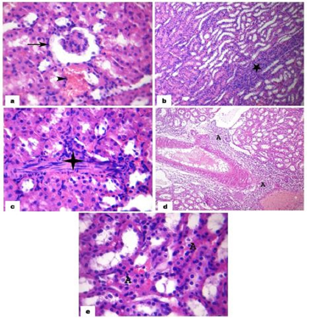 Fig. 1. Photomicrograph of rat kidney section stained with H&E of lead group (a,b,c,d) showing shrunken glomeruli with widening of glomerular space (arrow) and congestion of interstitial blood vessel (arrow head) (a) (×400), wide area of renal tubular necrosis with mononuclear inflammatory cells infilteration (star) (b) (×200), renal tubular necrosis (A) and fibrosis (asterisk) (c) (×400) with wide spread perivascular mononuclar inflammatory cells infilteration (A) (d) (×200) and a section in a rat kidney of lead+lycopene group showing minimal renal tubular necrosis (A) (e) (×400). Table 3. The score of detected renal lesions in male Wistar albino rats of different experimental groups
1Number of rats with lesions per total examined (7 rats). 2Severity of lesions was graded by estimating the percentage area affected in the entire section. |
|||||||||||||||||||||||||||||||||||||||||||||||||||||||||||||||||||||||||||||||||||||||||||||||||||||||||||||||||||||||||||||||||||||||||||||||||||||||||||
|
|
|||||||||||||||||||||||||||||||||||||||||||||||||||||||||||||||||||||||||||||||||||||||||||||||||||||||||||||||||||||||||||||||||||||||||||||||||||||||||||
|
Discussion |
|||||||||||||||||||||||||||||||||||||||||||||||||||||||||||||||||||||||||||||||||||||||||||||||||||||||||||||||||||||||||||||||||||||||||||||||||||||||||||
|
Lead is a common industrial and environmental heavy metal or pollutant that has been proved to be present in all phases of environment and biological systems. The accumulation of lead in the animals and human bodies and its related health risk became an issue of current debate and interest (Juberg et al., 1997). Liver, Kidney and brain are the main target organs for lead to express its toxic effect (Sharma and Street, 1980), which depends on ROS generation (Patra et al., 2001) but, the maximum accumulation of this biotoxicant is reported in the kidneys upon chronic exposure (Humphreys, 1991). So, this study aimed to evaluate the protective effect of lycopene (as a natural dietary antioxidant) against lead-induced nephrotoxicity. Lead-related nephrotoxicity is mainly associated with lipid peroxide formation due to ROS generation, which affects mitochondrial functions causing its damage and a subsequent cellular death (Wang et al., 2010a; Sen et al., 2006). The previously explained mechanism may demonstrate the significant increase in serum concentration of urea and creatinine of Pb-intoxicated rats which indicated the decrease in glomerular filtration rate (due to glomerular damage) of these animals as a consequence of Pb intoxication (Jayakumaret al., 2009; Wang et al., 2010b; Wang et al., 2013). Combined with these findings, the concentration of serum cystatin-C was significantly increased in Pb-intoxicated rats and this may affirm the nephrotoxic effect of lead as cystatin-C is a non-glycosylated protein, which is fully catabolized by renal tubules after filtration without any reabsorption, so it is considered as a more accurate marker of renal filtration power (Murty et al., 2013). In parallel, AKIM-1 is a transmembrane protein, which is highly upregulated in tubular epithelium after kidney injury, shed into tubular lumen and reabsorbed to reach blood, so it is one of the most accurate blood biomarker for tubular injury (Sabbisetti et al., 2014) and this may demonstrate its significant elevation in serum of Pb-intoxicated group. The elevation in the previously discussed nephrotoxic biomarkers effectively proved both of glomerular and tubular injuries caused by lead intoxication. Oxidative stress evoked by lead intoxication was declared in this study by the significant increase in renal tissues concentration of lipid peroxide (MDA), which coupled with significant depletion of enzymatic antioxidants (GSH, SOD and CAT) (Patra et al., 2001; Sivaprasad et al., 2002; Wang et al., 2010b; Wang et al., 2013). Lycopene is one of the most powerful antioxidant among different known carotenoids (Hazewindus et al., 2012) and this could be a reason for the potential role of lycopene in partial recovery of biochemical and oxidant-antioxidant alteration related to lead peroxidative nephrotoxicity, particularly by reducing oxidative stress in renal tissues as recorded by several studies (Ateşşahin et al., 2005; Ateşşahin et al., 2007). Experimental and clinical trials have indicated that the inflammatory process mediated by innate immune responses plays a crucial role in the development and progression of lead nephrotoxicity as the levels of the pro-inflammatory cytokines specially, TNF-α, IL-6, and IL-1β were increased in renal tissues of lead-intoxicated rats (Liu et al., 2012; Salama et al., 2016). In the present study, the increment in serum level of these pro-inflammatory cytokines was mitigated upon lycopene treatment which suggests that lycopene has an important implication in anti-inflammatory reaction (Di Mascio et al., 1989; Bignotto et al., 2012; Hadad and Levy, 2012). Pyruvate kinase enzyme is the master enzyme controlling glycolysis in most of the body tissues as it catalyzes the final step of glycolysis to yield ATP (Gupta and Bamezi, 2010). Findings from this study declared that PK enzyme activity in renal tissues of lead-intoxicated rats was significantly decreased which may have a role in occurrence of renal cellular death due to energy deprivation in case of lead intoxication. The reason may be that PK contains a sulfhydryl group, which is important for its properties and catalytic activity (Tomich and Colman, 1985; Rafter and Blair, 1987) and unfortunately, lead is known as an inhibitor for sulfhydryl containing enzymes (Vallee and Ulmer, 1972; Gurer et al., 1999) including PK enzyme (Yun and Hoyer, 2000; Lepper et al., 2010). On the other hand, G-6-P enzyme can liberate glucose from glucose-6-phosphate upon cellular need, but it was detected that the destruction of microsomal G-6-P enzyme may occur as a result of exposure to lipid peroxide (Hruszkewycz et al., 1978; Daniels et al., 1995) and this may explain the decrease in renal activity of this enzyme in lead intoxicated animals (due to lead-related oxidative stress). Fortunately, the previously affirmed antioxidant effect of lycopene may assist in amelioration of PK enzymes inhibition through reservation of renal GSH level as GSH has a great role as a chelator for heavy metals including lead (Reed, 1990; Meister, 1994) decreasing its effect on thiol containing enzymes as PK. In addition, lycopene may alleviate the destruction of G-6-P enzyme via relieving of lead-induced oxidative stress (ROS generation) and its resultant lipid peroxide which inhibit G-6-P enzyme. Histopathologically, lead-related oxidative nephrotoxicity is characterized by glomerular sclerosis, proximal tubular nephropathy and interstitial fibrosis (Loghman-Adham, 1997; Diamond, 2004) and this came in a total accordance with our results concerning histopathological evaluation of lead intoxicated rat's kidneys, also, the histopathological findings confirmed the obtained biochemical results and the ameliorative role of lycopene against lead-induced nephrotoxicity. |
|||||||||||||||||||||||||||||||||||||||||||||||||||||||||||||||||||||||||||||||||||||||||||||||||||||||||||||||||||||||||||||||||||||||||||||||||||||||||||
|
|
|||||||||||||||||||||||||||||||||||||||||||||||||||||||||||||||||||||||||||||||||||||||||||||||||||||||||||||||||||||||||||||||||||||||||||||||||||||||||||
|
Conclusion |
|||||||||||||||||||||||||||||||||||||||||||||||||||||||||||||||||||||||||||||||||||||||||||||||||||||||||||||||||||||||||||||||||||||||||||||||||||||||||||
|
This study illuminated the inhibitory effect of lead intoxication-related oxidative stress on PK and G-6-P enzymes in renal tissues as a co-factor for occurrence of renal cellular injury and proved the attenuative role of lycopene as an antioxidant and anti-inflammatory substance in mitigation of lead-related nephrotoxicity. |
|||||||||||||||||||||||||||||||||||||||||||||||||||||||||||||||||||||||||||||||||||||||||||||||||||||||||||||||||||||||||||||||||||||||||||||||||||||||||||
|
|
|||||||||||||||||||||||||||||||||||||||||||||||||||||||||||||||||||||||||||||||||||||||||||||||||||||||||||||||||||||||||||||||||||||||||||||||||||||||||||
|
Conflict of Interests |
|||||||||||||||||||||||||||||||||||||||||||||||||||||||||||||||||||||||||||||||||||||||||||||||||||||||||||||||||||||||||||||||||||||||||||||||||||||||||||
|
The author declared no conflict of interests exists. |
|||||||||||||||||||||||||||||||||||||||||||||||||||||||||||||||||||||||||||||||||||||||||||||||||||||||||||||||||||||||||||||||||||||||||||||||||||||||||||
|
|
|||||||||||||||||||||||||||||||||||||||||||||||||||||||||||||||||||||||||||||||||||||||||||||||||||||||||||||||||||||||||||||||||||||||||||||||||||||||||||
|
References |
|||||||||||||||||||||||||||||||||||||||||||||||||||||||||||||||||||||||||||||||||||||||||||||||||||||||||||||||||||||||||||||||||||||||||||||||||||||||||||
|
Abdel Moneim, A.E., Dkhil, M.A., Al-Quraishy, S., 2011. The protective effect of flaxseed oil on lead acetate-induced renal toxicity in rats. J. Hazard. Mater. 194, 250–255. Abdel-Moneim, A.E., Dkhil, M.A, Al-Quraishy, S., 2011. The redox status in rats treated with flaxseed oil and lead-induced hepatotoxicity. Biol. Trace. Elem. Res. 143, 457–467. Ateşşahin, A., Çeribaş, A.O., Yılmaz, S., 2007. Lycopene, a carotenoid, attenuates cyclosporine-induced renal dysfunction and oxidative stress in rats. Basic Clin. Pharmacol. Toxicol. 100, 372–376. Ateşşahin, A., Yilmaz, S., Karahan, I., Ceribasi, A.O., Karaoglu, A., 2005. Effects of lycopene against cisplatin-induced nephrotoxicity and oxidative stress in rats. Toxicol. 212, 116–123. Augusti, P.R., Conterato, G.M., Somacal, S., Einsfeld, L., Ramos A.T., Hosomi, F.Y., Emanuelli T., 2007. Effect of lycopene on nephrotoxicity induced by mercuric chloride in rats. Basic Clin. Pharmacol. Toxicol. 100, 398–402. Bancroft, J.D., Stevens, A., 1996. Theory and Practice of Histological Technique. Fourth Ed. Churchil, livingestone, USA. Bignotto, L., Rocha, J., Sepodes, B., Eduardo-Figueira, M., Pinto, R., Chaud, M., Carvalho, J., Moreno, H., Mota-Filipe, H., 2012. Anti-inflammatory effect of lycopene on carrageenan-induced paw oedema and hepatic ischaemia–reperfusion in the rat. Br. J. Nutri. 102, 126–133. Chang, L.W., Wade, P.R., Olson, M.N., 1980. Ultrastructural changes in renal proximal tubules after tetraethyle lead intoxication. Environ. Res. 23, 208–223. Courtois, E., Marques, M., Barrientos, A., 2003. Lead-induced down regulation of soluble guanylate cyclase in isolated rat aortic segments mediated by reactive oxygen species and cyclooxygenase-2. J. Am. Soc. Nephrol. 14, 1464–1470. Daniels, W.M., Reiter, R.J., Melchiorri D, Sewerynek, E., Pablos, M.I., Ortiz, G.G., 1995. Melatonin counteracts lipid peroxidation induced by carbon tetrachloride but does not restore glucose 6 phosphatase activity. J. Pineal. Res. 19, 1-6. Di Mascio, P., Kaiser, S., Sies, H., 1989. Lycopene as the most efficient biological carotenoid singlet oxygen quencher. Arch. Biochem. Biophys. 274, 532–538. Diamond, G.L., 2004. Risk assessment of nephrotoxic metals. In: Tarloff J and Lash L (Eds) The Toxicology of the Kidney. CRC Press, London, England, pp. 1099–1132. El-Sokkary, G.H., Abdel-Rahman, G.H., Kamel, E.S., 2005. Melatonin protects against lead-induced hepatic and renal toxicity in male rats. Toxicol. 213, 25–33. Ercal, N., Treeratphan, P., Hammond, T.C., Matthews, R.H., Grannemann, N.H., Spitz, D.R., 1996. In vivo indices of oxidative stress in lead-exposed c57bl/6 mice are reduced by treatment with meso- 2, 3-dimercaptosuccinic acid or n-acetylcysteine. Free Radic. Biol. Med. 21, 157–161. Gupta, V., Bamezi, R.N., 2010. Human Pyruvate kinase M2: A multifunctional protein. Protein. Sci. 19, 2031–2044. Gurer, H., Ercal, N., 2000. Can antioxidants be beneficial in the treatment of lead poisoning? Free. Radic. Biol. Med. 29, 927–945. Gurer, H., Neal, R., Yang, P., Oztezcan, S., Ercal, N., 1999. Captropril as an antioxidant in lead-exposed Fischer 344 rats. Human Exper. Toxicol. 18, 27–32. Hadad, N., Levy, R., 2012. The synergistic anti-inflammatory effects of lycopene, lutein, β-carotene, and carnosic acid combinations via redox-based inhibition of NF-κB signaling. Free. Radical Biol. Med. 53, 1381–1391. Halliwell, B., Murcia, M.A., Chirico, S., Aruoma, O.I., 1995. Free radicals and antioxidants in food and in vivo: what they do and how they work. Crit. Rev. Food. Sci. Nutr. 35, 7–20. Halliwell, B., 1994. Free radicals, antioxidants and human disease: Curiosity, cause or consequence? Lancet 344, 721-724. Hazewindus, M., Haenen, G.R., Weseler, A.R., Bast, A., 2012. The anti-inflammatory effect of lycopene complements the antioxidant action of ascorbic acid and α-tocopherol. Food Chem. 132, 954–958. Hruszkewycz, A.M., Glende-Jr, E.A., Recknagel, R.O., 1978. Destruction of microsomal Cytochrome p450 and Glucose-6-phosphatase by lipids extracted from peroxidized microsomes. Toxicol. App. Pharmacol. 46, 695–702. Humphreys, D,J., 1991. Effect of exposure to excessive quantities of lead animals. Br Vet. J. 147, 18–30. Jayakumar, T., Sridhar, M.P., Bharathprasad, T.R., Ilayaraja, M., Govindasamy, S., Balasubramanian, M.P., 2009. Experimental studies of Achyranthes aspera (L) preventing nephrotoxicity induced by lead in albino rats. J. Health. Sci. 55, 701–708. Juberg, D.R., Klieman, C.F., Kwon, S.C., 1997. Position paper of the American Council on Science and Health: Lead and human health. Ecotoxicol. and Environmental Safety 38, 162–180. Khalil-Manesh, F., Gonick, H.C., Cohen, A.H., Alinovi, R., Bergamaschi, E., Mutti, A., Rosen, V.J., 1992. Experimental model of lead nephropathy. I. Continuous high dose lead administration. Kidney Int. 41, 1192–1203. Lepper, T.W., Oliveira, E., Koch, G.D.W., Berlese, D.B., Feksa, L.R., 2010. Lead inhibits in vitro creatine kinase and pyruvate kinase activity in brain cortex of rats. Toxicol. in Vitro 24, 1045–1051. Lin, J.L., Lin-Tan, D.T., Li, Y.J., Chen, K.H., Huang, Y.L., 2006. Low-level environmental exposure to lead and progressive chronic kidney diseases. Am. J. Med. 119, 707. Lindshield, B.L., Canene-Adams, K., Erdman, J.W., 2007. Lycopenoids: are Lycopene Metabolites Bioactive?. Arch. Biochem. Biophys. 458, 136–140. Liu, C.M., Sun, Y.Z., Sun, J.M., Ma, J.Q., Cheng, C., 2012. Protective role of quercetin against lead-induced inflammatory response in rat kidney through the ROS-mediated MAPKs and NF-κB pathway. Biochimica. et Biophysica. Acta (BBA)-General Subjects 1820, 1693–1703. Loghman-Adham, M., 1997. Renal effects of environmental and occupational lead exposure Environ. Health Perspect. 105, 928–939. Mahalley, K.R., 1977. Relation between quantities of lead ingested and health effects of lead in humans. Pediatrics 59, 448–456. Meister, A., 1994. Glutathione, ascorbate, and cellular protection. Cancer Res. 54, 1969–1975. Missoun, F., Slimani, M., Aoues, A., 2010. Toxic effect of lead on kidney function in rat Wistar. Africa J. Biochem. Res. 4, 21–27. Murty, M.S., Sharma, U.K., Pandey, V.B., Kankare, S.B., 2013. Serum cystatin C as a marker of renal function in detection of early acute kidney injury. Indian J. Nephrol. 23, 180–183. Patra, R.C., Rautray, A.K., Swarup, D., 2011. Oxidative stress in lead and cadmium toxicity and its amelioration. Vet. Med. Int. 2011. Patra, R.C., Swarup, D., Dwivedi, S.K., 2001. Antioxidant effects of tocopherol, ascorbic acid and L-methionine on lead induced oxidative stress to the liver, kidney and brain in rats. Toxicol. 162, 81–88. Rafter, G.W., Blair, J.B., 1987. Reaction of liver pyruvate kinase with sulfhydryl reagents: effect on its activity. Biochim. Biophys. Acta 913, 195–199. Rao, A.V., Agarwal, S., 1999. Role of Lycopene as Antioxidant Carotenoid in The Prevention of chronic Diseases: A Review. Nutri. Res. 19 (2), 305–323. Rao, A.V., Agarwal, S., 2000. Tomato lycopene and its role in human health and chronic diseases. CMAJ 19, 163–166. Reed, D.J., 1990. Glutathione: toxicological implications. Ann. Rev. Pharmacol. Toxicol. 30, 603–631. Sabbisetti, V.S., Waiker, S.S., Antoine, D.J., Smiles, A., Wang, C., 2014. Blood kidney injury molecule-1 is a biomarker of acute and chronic kidney injury and predict progression to ESRD in type I diabetes. J. AM. Soc. Nephrol. 25, 2177–2186. Salama, S.A., Arab, H.H., Maghrabi, I.A., Hassan, M.H., Al-Saeed, M.S., 2016. Gamma-Glutamyl Cysteine Attenuates Tissue Damage and Enhances Tissue Regeneration in a rat Model of Lead-Induced Nephrotoxicity. Biolo. Trace Element Res. 173, 96–107. Sen, T., Sen, N., Tripathi, G., Chatterjee, U., Chakrabarti, S., 2006. Lipid peroxidation associated cardiolipin loss and membrane depolarization in rat brain mitochondria. Neurochem. Int. 49, 20–27. Sharma, R.P., Street, J.C., 1980. Public health aspects of toxic heavy metals in animal feeds. J. Am. Vet. Med. Assoc. 177, 149–153. Sies, H., Stahl, W., 1995. Vitamins E and C, beta-carotene, and other carotenoids as antioxidants. Am. J. Clin. Nutr. 62, 1315–1321. Sivaprasad, R., Nagaraj, M., Varalakashmi P., 2002. Lipoic acid in combination with chelator ameliorate lead-induced peroxidative damage in rat kidney. Arch. Toxicol. 76, 437–441. Smith, D.R., Kahng, M.W., Quintanilla-Vega, B., Fowler, B.A., 1998. High-affinity renal lead-binding proteins in environmentally-exposed humans. Chem. Biol. Interact. 115, 39–52. Swanson, M.A., 1955. Glucose-6-phosphatase from liver. In: Methods in Enzymology (Colowick, S.P., Kaplan, N.O., Eds). Academic Press, New York, pp. 541–543. Tomich, J.M., Colman, R.F., 1985. Reaction of 59-p-fluorosulfonylbenzoyl-1, N6- ethanoladenosine with histidine and cysteine residues in the active site of rabbit muscle pyruvate kinase. Biochim. Biophys. Acta 827, 344–357. Vallee, B.L., Ulmer, D.D., 1972. Biochemical effect of mercury, cadmium and lead. Ann. Rev. Biochem. 41, 91–115. Wang, L., Li, J., Li, J., Liu, Z., 2010a. Effects of lead and/or cadmium on the oxidative damage of rat kidney cortex mitochondria. Biol. Trace Elem. Res. 137, 69–78. Wang, L., Lin, S., Li, Z., Yang, D., Wang, Z., 2013. Protective effects of puerarin on experimental chronic lead nephrotoxicity in immature female rats. Human Exper. Toxicol. 32, 172–185. Wang, L., Wang, Z., Liu, J., 2010b. Protective effect of N-acetylcysteine on experimental chronic lead nephrotoxicity in immature female rats. Human Exper. Toxicol. 29, 581–591. Yilmaz, S., Atessahin, A., Sahna, E., Karahan, I., Ozer, S., 2006. Protective effect of lycopene on adriamycin-induced cardiotoxicity and nephrotoxicity. Toxicol. 218, 164–171. Yun, S.W., Hoyer, S., 2000. Effects of low-level lead on glycolytic enzymes and pyruvate dehydrogenase of rat brain in vitro: relevance to sporadic Alzheimer’s disease? J. Neural. Trans. 107, 355–368. |
|||||||||||||||||||||||||||||||||||||||||||||||||||||||||||||||||||||||||||||||||||||||||||||||||||||||||||||||||||||||||||||||||||||||||||||||||||||||||||
|
|
|||||||||||||||||||||||||||||||||||||||||||||||||||||||||||||||||||||||||||||||||||||||||||||||||||||||||||||||||||||||||||||||||||||||||||||||||||||||||||