|
|
Journal of Advanced Veterinary Research Volume 9, Issue 3, 2019, Pages: 128-129 www.advetresearch.com |
|


|
Management of Fetal dystocia due to Downward Deviation of Head with Bilateral Carpal Flexion in a non-descript Local Goat |
|
|
|
Asloob Ahmad Malik*, Bilal Ahmad Ganaie, Rayees Habib, Muneeb Shabir |
|
|
|
Division of Animal Reproduction, Gynaecology and Obstetrics, Faculty of Veterinary Sciences and Animal Husbandry, Sher-e-Kashmir University of Agricultural Sciences & Technology of Kashmir, Shuhama Alusteng, Srinagar – 190006, India. |
|
|
|
Received: 21 June 2019; Accepted: 11 July 2019 |
|
*Corresponding author: Asloob Ahmad Malik (e-mail: malikasloob@gmail.com) |
|
Abstract |
|
A case of dystocia in a local non-descript goat was reported. The animal was weak and had a history of straining since 24 hours, fetal membranes ruptured and cervix fully dilated. On per-vaginal examination the fetus was found dead as there was no reflex on pinching. The fetus had a normal presentation, position but posture was abnormal, with downward deviation of head and both the carpal joints were flexed. Caudal epidural anaesthesia was given between first and second intercoccygeal space using 2.5 ml Lignocaine hydrochloride before manual handling. After proper lubrication, correction of the dystocia was carried out using the repulsion and traction method. Antibiotics and anti-inflammatory were injected post successful traction of the fetus. Then, 500 ml of 5% Dextrose fluid was administered intravenously to correct the dehydration status and to avoid shock to the animal. After successful traction of fetus, two furea boli were placed intrauterine. |
|
|
|
Keywords: Bilateral carpel flexion, Dystocia, Downward deviation of head, Presentation |
|
|
|
Introduction |
|
|
|
Dystocia refers to the condition in which animal fails to expel the fetus at the time of parturition and human intervention becomes a necessity (Blood et al., 2011). The incidence of dystocia in small ruminants is very low worldwide averaging around 5 per cent (Sharma et al., 2014; Bhattacharyya et al., 2015). The causes of dystocia in goats have been broadly classified into two categories namely fetal causes- including fetal malpresentation and malposition, oversized fetus and congenital abnormalities like hydrocephalic condition. While as maternal causes include uterine inertia having multiple foetuses, over feeding of dam during pregnancy and narrow pelvis (Pugh and Baird, 2012). The chances of fetal and maternal losses increase with the delay in external intervention. It has also been reported that with prolonged dystocia the chances of necrotic metritis are high which usually proves fatal (Scott, 2006). Dystocia induced trauma and infection reduces future fertility of the animal (Aziz and Taha, 1996). This clinical case report presents the management of dystocia in a local goat caused by downward deviation of head with bilateral carpel flexion. |
|
|
|
Case history and clinical observation An adult female goat weighing 35-40 kg was presented to the Teaching Veterinary Clinical Complex, Faculty of Veterinary Sciences and Animal Husbandry, Shuhama Alusteng, with the history of straining since 24 hours and difficulty in giving birth. The deworming status was up-to-date. Physical examination of the animal revealed pale mucous membrane with capillary refill time of 4 sec. The goat was weak and the fetal membranes had already ruptured with none of the fetal parts visible at the vulval region, however the cervix was fully dilated. Per-vaginal examination revealed that the fetus was already dead as there was not any sort of reflex on pinching. However, the fetal presentation was found as normal with anterior presentation, normal dorso-sacral position, but posture was abnormal where along with downward deviation of head both carpal joints were flexed. This posture had resulted in the engagement of the fetal mane region in the pelvic brim of the dam. Therefore, based on pervaginal examination, the condition was diagnosed as fetal dystocia due to downward deviation of head and bilateral carpal flexion posture. |
|
|
|
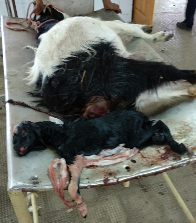
Treatment and Discussion The treatment plan for present case was to remove the dead fetus via mutation and traction method. Caudal epidural anaesthesia was given between first and second inter-coccygeal space using 2.5 ml Lignocaine hydrochloride (Lox 2%, Neon Laboratories Ltd, India) before the procedure. Liquid paraffin solution was used to lubricate the birth canal so as to facilitate easy manipulation of the fetus. After proper lubrication, correction of the dystocia was carried out using the repulsion and traction method. The limbs were identified and carpel flexion was corrected by cupping the hooves and bringing the limbs into the birth passage. After correction of the carpel flexion, both the limbs were pushed back into the uterine cavity to create working space for correction of fetal head. The downward deviation of head was corrected after eye hook was inserted in the medial canthus of the eye orbit and traction was applied slowly till the head was brought in the vaginal passage. Finally, the limbs were retracted and traction was applied on both limbs and a male dead fetus was successfully delivered (Fig. 1). Dexona Vet 5ml (Zydus AHL, India) and Revici 3ml (Kee Pharma, India) were administered intramuscularly. This was followed by Levofloxacin 3ml (Meriflox; Vetoquinol Ltd, India) and 2.5ml Meloxicam (Melonex, Intas Pharma Ltd, India). Then, 500 ml of 5% Dextrose fluid was administered intravenously to correct the dehydration status and to avoid shock to the animal. After successful traction of fetus, two Furea boli (Pfizer Ltd, India) were placed intrauterine. The animal had an uneventful recovery thereafter.
Fig 1. Male dead fetus lying alongside of the Goat |
|
|
|
References |
|
Aziz, D.M., Taha, M.B., 1996. Dystocia in Awassi ewes: causes and treatments a review. Iraqi Journal of Veterinary Science 9, 1-12. Bhattacharyya, H.K., Fazili, M.R., Bhat, F.A., Buchoo, B.A., 2015. Prevalence and dystocia of sheep and goats: A study of 70 cases (2004-2011). Journal of Advanced. Veterinary Research 5, 14-20. Blood, D.C., Studdert, V.P., Gay, C.C., 2011. Saunders Comprehensive Veterinary Dictionary 4th Edn; London: Saunders. Pugh, D.G., Baird, N.N., 2012. Sheep and goat medicine. Elsevier Health Sciences. Scott, P.R., 2006. Sheep Medicine. Manson Publishing. Sharma, A., Kumar, P., Singh, M., Vasishta, N., 2014. Retrospective analysis of dystocia in small ruminants. Intas Polivet. 15, 287-89. |
|
|