|
|
Journal of Advanced Veterinary Research Volume 10, Issue 1, 2020, Pages: 17-20 www.advetresearch.com |
|


|
Evaluation of Testicular Blood Flow and Ultrasonographic Measurements in Rams with Emphasis on Laterality |
|||||||||||||||||||||
|
|
|||||||||||||||||||||
|
Mohamed G. Hedia1*, Mohamed S. El-Belely1, Sayed T. Ismail1, Amal M. Abo El-Maaty2 |
|||||||||||||||||||||
|
1Department of Theriogenology, Faculty of Veterinary Medicine, Cairo university, Giza, Egypt. 2Department of Animal Reproduction and AI, Veterinary Division, National Research Center, Dokki, Giza, Egypt. |
|||||||||||||||||||||
|
Received: 4 December 2019; Accepted: 27 December 2019 (*: Corresponding author: mohammedhedia@cu.edu.eg) |
|||||||||||||||||||||
|
|
|||||||||||||||||||||
|
Abstract |
|||||||||||||||||||||
|
|
|||||||||||||||||||||
|
The current study aimed to measure the testicular ultrasonographic dimensions (length, width, thickness and volume) as well as spectral Doppler indices (resistive index and pulsatility index) of testicular artery, and to investigate the possible differences between the paired testes based on testicular dimensions and blood flow Doppler indices. Five Awassi rams of normal fertility, with a body weight 50-65 kg and aged 3-5 years were subjected to testicular ultrasound examination for testicular dimensions and pulsed-wave Doppler, twice monthly for five consecutive months. The testicular volume of the left testis (69.34±6.66 mm3) was higher than the right testis (66.85±5.46 mm3). Testicular thickness was significantly affected by the laterality of the testes (P<0.05). Doppler measures for RI (resistive index) as well as pulsatility index (PI) were slightly higher (P>0.05) in the right testicular artery rather than the left one. In conclusion, the testicular ultrasound measures and Doppler flow indices varied between the paired right and left testes in mature rams. In addition, the blood flow indices are highly correlated in the testicular artery of rams. |
|||||||||||||||||||||
|
|
|||||||||||||||||||||
|
Keywords: Testes, Doppler, Ultrasound, Rams, Laterality |
|||||||||||||||||||||
|
|
|||||||||||||||||||||
|
Introduction |
|||||||||||||||||||||
|
|
|||||||||||||||||||||
|
The testis is a paired organ and considered as the main reproductive component in the male, which is responsible for both exocrine through gamete production as well as testosterone formation (Setchell, 1978). Ultrasonographic Doppler imaging is widely used as trans-abdominal and trans-rectal diagnostic imaging (Rubens et al., 2006) owing to its several advantages as a safe, noninvasive method to evaluate testicular parenchyma in rams (Camela et al., 2019). For maximal meat and milk production, producers tend to house a limited number of rams with a peak reproductive efficiency, which allow them to impregnate a large flock of ewes in the same breeding system. Breeding soundness examination (BSE) is a series of definite steps, which are done to indirectly assess the reproductive performance of rams through evaluation of certain physical, reproductive and ultrasonographical parameters, which is mainly through measuring the testicular dimensions for length, width, thickness and volume of testicular parenchyma as well as pulsed-wave Doppler indices, as resistive index and pulsatility index (Batissaco et al., 2013; Camela et al., 2019). There are different measures of blood flow velocities and spectral Doppler indices of the testicular artery, which could be evaluated using pulsed-wave Doppler ultrasonography. Peak systolic velocity (PSV), end diastolic velocity (EDV), mean velocity (MV), resistive index (RI) and pulsatility index (PI). Interestingly, measures of blood flow velocities within the arterial blood vessel during the heart cycle are different and not stable between measurements (Pozor and McDonnell, 2004). The testicular artery supplies a stable blood flow needed for the metabolic process and spermatogenesis (Bergh and Damber, 1993). The RI (a measure of blood flow that reflects the resistance to blood flow caused by microvascular beds distal to the site of measurement) and PI (conducted to quantify the pulsatility of oscillations of the waveform) are vastly used to study testicular blood flow dynamics in human and animals (Biagiotti et al., 2002; Batissaco et al., 2013; de Souza et al., 2014). As far as we know, there are limited reports concerning the use of testicular ultrasonography to assess the reproductive capacity of Awassi rams in Egypt. On the other hand, the influence of testicular laterality on fluctuations of testicular dimensions using grey-scale ultrasonography as well as assessment of testicular blood irrigation using pulsed-wave Doppler has to be recorded in Awassi rams under Egyptian subtropical conditions. Thus, the current study aimed to record the definite values of testicular dimensions and volume from one hand, and to measure the blood flow indices of the testicular artery from the other hand, for right and left testes in rams. |
|||||||||||||||||||||
|
|
|||||||||||||||||||||
|
Materials and methods |
|||||||||||||||||||||
|
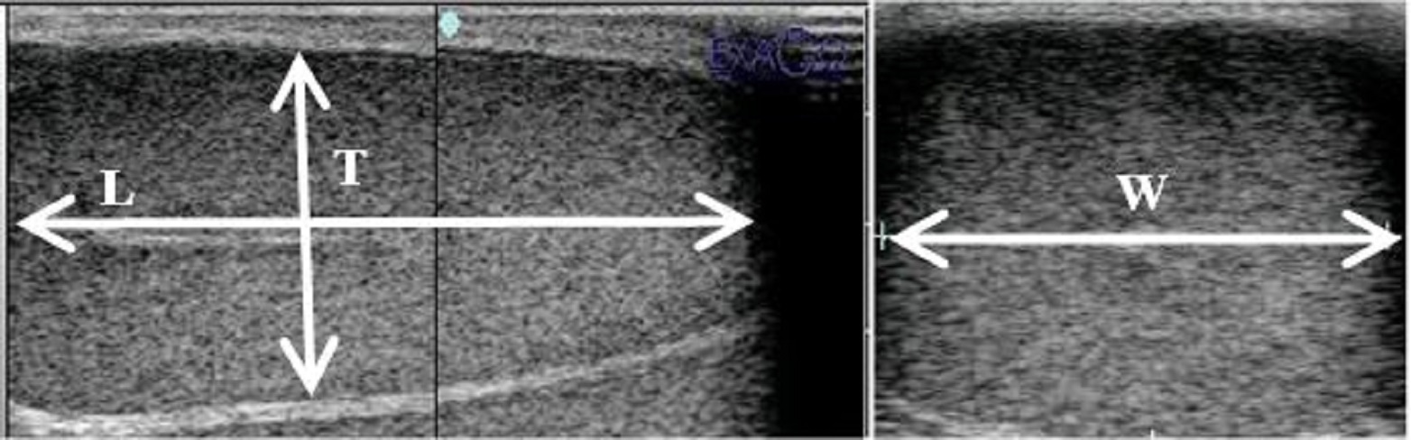
This protocol was performed at Theriogenology Department, Faculty of Veterinary Medicine, Cairo University, Egypt. The experimental protocol was approved by the Ethics Committee of Cairo University (IACUC). The present study was carried out for five successive months, where each examination trial was conducted twice monthly. Animals and management Five mature Awassi rams of normal reproductive performance, weighing 50-65 kg and aging 3-5 years old were kept under natural macro-environment (ambient temperature and daylight). In accordance with NRC recommendations (NRC, 2007), each ram fed daily 1.25 kg ration composed of 850 g roughages and green forage and 400 g pelleted concentrates with free access to fresh water. Rams were routinely vaccinated against the endemic diseases and, and intensively checked for both external and internal parasites. Importantly, the circulatory system was checked to get rid of the ram suffering from any cardiovascular problem that influencing the testicular blood flow measurements. All selected rams showed normal ranges of pulse rate (60-90 beat/min.) and capillary refilling time test (1-2 s) during the whole length of the study period (Pugh and Baird, 2012). Ultrasound examinations Scanning was carried out using ultrasound scanner (EXAGO, Echo Control Medical, France) equipped with a linear array transducer (7.5 MHz). The same investigator evaluated the Doppler ultrasonographic measurements during the whole study. Estimating testicular dimensions by B- mode ultrasonography The rams were secured without sedation in the lateral recumbency position. Before any examination procedure, the fine wool at the both sides of the scrotum were completely removed, to ensure the direct contact between ultrasound transducer and the scrotal skin. After that the transducer was covered with adequate amount of gel to eliminate the presence of air spaces to facilitate the ultrasonographic scanning. For proper examination, three separate transverse and longitudinal images of each testis were measured, and the testicular length (L), thickness (T) and width (W) were recorded using electronic callipers (Fig. 1). The testis volume was calculated using the formula L × T × W × 0.71 (Raji et al., 2016).
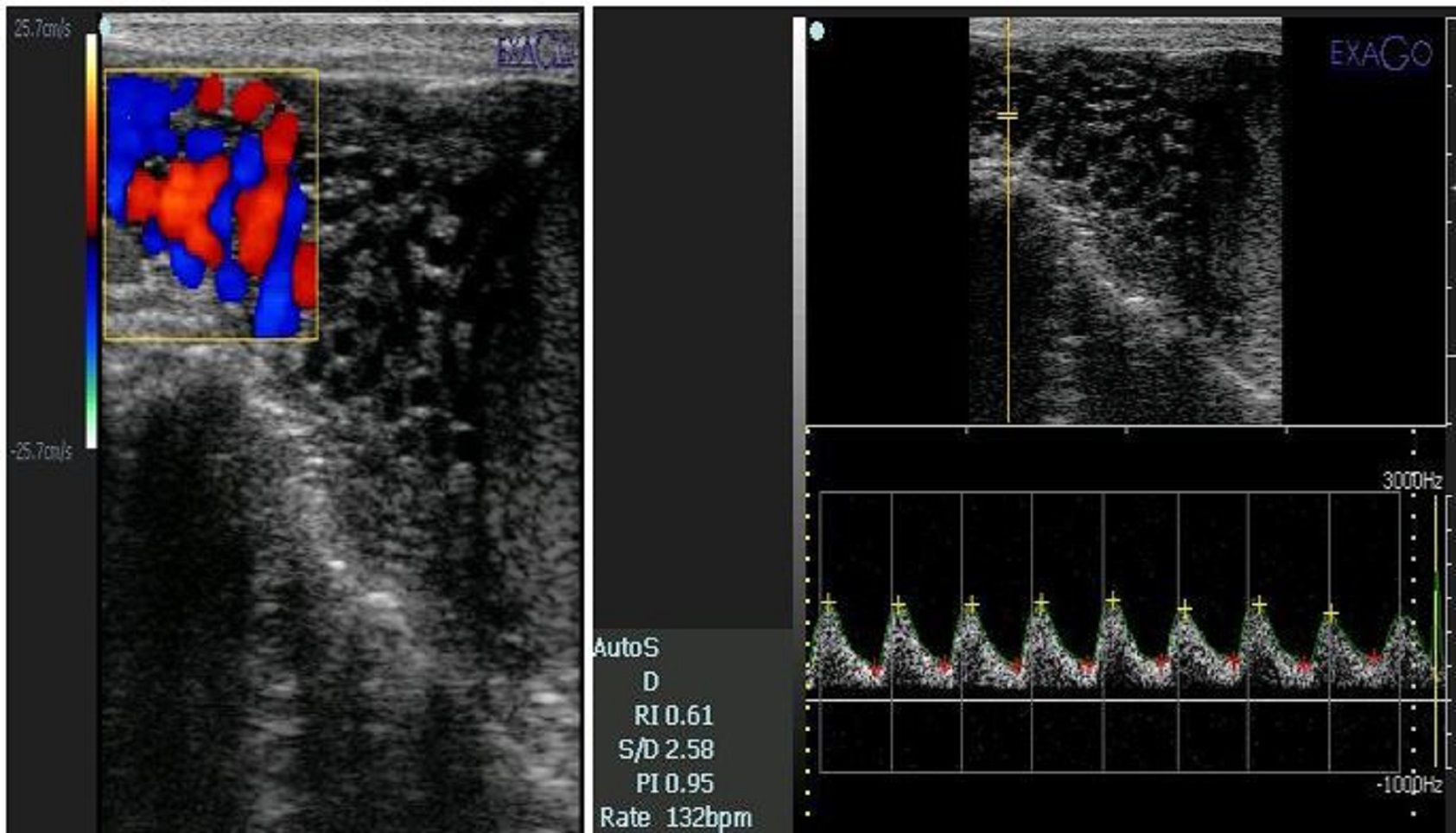
Fig. 1. Testis of fat-tailed ram imaged by B-mode ultrasonography for measurement of testicular volume according to the formula: L×T×W×0.71 (L= length, T= thickness and W= width). Measurements of the testicular artery using Doppler After locating the colored box of blood flow at the proximal aspect of the testis using the color mapping mode. Investigator recorded Three to five measurements for each parameter in different locations along the path of the testicular artery. The ultrasound settings including focus, gains, brightness and contrast were standardized, stabilized and used equally for all examinations. Interestingly, the angle between the Doppler beam and the long axis of the vessel was kept between 45- 60 in the direction of blood flow with the high pass filter set at 50 Hz. The Doppler gate was fixed for all scanning trials at 1 mm. After locating the testicular artery, investigator fixed the examination box through the arterial axis, and recorded the spectral pattern of the testicular artery using the pulsed-wave mode (Fig. 2), the measured Doppler indices were resistive index (RI = (maximum velocity - minimum velocity)/maximum velocity) and pulsatility index (PI = (maximum velocity-minimum velocity)/mean velocity) according to Batissaco et al. (2013).
Fig. 2. Ultrasonographic scanning of the fat-tailed ram testis using color pulsed-wave Doppler ultrasonography showing blood flow within the testicular artery (left side). Blood flow within the supratesticular artery revealed a spectral pattern for measurement of blood flow indices within the testicular artery (right side). Statistical analysis The data were tested for normality (Shapiro-Wilk test). Descriptive analysis for the studied variables was expressed as mean ±SEM. An analysis of variance (ANOVA) was performed for the comparisons between right and left testes using paired t test (Steel and Torrie, 1980). Pearson correlation coefficients were checked among the studied variables. A significance level of P <0.05 was used in all cases. All data were statistically analyzed through the software SPSS (Statistical Package for Social Sciences). |
|||||||||||||||||||||
|
|
|||||||||||||||||||||
|
Results |
|||||||||||||||||||||
|
Descriptive statistics for testicular dimensions as well as blood flow through testicular artery are shown in Table 1. Testicular length, resistive index as well as pulsatility index showed none significant changes for the right testis compared to the left one. Results demonstrated that there was a significant influence of testicular laterality on testicular thickness (P<0.05). However, no considerable changes were found between the other measured variables (P>0.05). Table 1. Summary of morphometric characteristics of the testes as well as velocimetric indices of the testicular artery (mean ±SEM) in rams.
Data were expressed as Mean±SE, *: Significant at P<0.05. Testicular thickness showed positive significant (P<0.01) associations with testicular length(r=0.350) and width (r=0.413), in addition, a higher positive (P<0.01) correlation was recorded between blood flow indices (r=0.0925). |
|||||||||||||||||||||
|
|
|||||||||||||||||||||
|
Discussion |
|||||||||||||||||||||
|
Intensive evaluation of testicular dimensions is of great potential benefit in the evaluation of testicular functionality. Since the testis is a highly compact organ, where the seminiferous tubules comprise 70-80% of testicular parenchyma, testicular volume is largely a reflection of sperm cells production (Sarlós et al., 2013). Testicular volume estimation is important to ensure the reproductive capacity. In addition, it can be a tool to observe pathologies that sustain in a testes alteration. Furthermore, the testicular volume can be a sign of infectious diseases or tumour in the organ, compromising the spermatogenesis, as it has reported in humans (Foresta et al., 1998; Shiraishi et al., 2009). Recently, ultrasonographic assessment of testicular dimensions known as the most accurate based upon the comparison with actual testicular volume in human beings and animals (Bollwein et al., 2008; Sakamoto et al., 2008; Zelli et al., 2013). The present results indicated that testicular size of left testis had a higher value than right one, which was confirmed previously in rams (Andrade et al., 2014; Focsaneanu et al., 2014; Karimi et al., 2019). The latter authors recorded a different average for testicular dimensions rather than those reported in the present study and the discrepancy might be due, in part, to the breed differences, geographical factors as well as the age of rams at time of evaluation. In addition, our results were also confirmed the same trend in other species as stallion (Kavak et al., 2003) and dogs (Souza et al., 2012). On pulsed-wave Doppler imaging, the testicular artery showed mostly monophasic and non-resistive waveforms pattern similar to that described in humans (Middleton et al., 1989), dogs (Zelli et al., 2013) as well as in rams (Batissaco et al., 2013). However, spectral Doppler imaging for the testicular artery in stallions exhibited resistive and biphasic waveforms (Pozor and McDonnell, 2004), these differences might be due, in part, to the vertical long axis of ram testes compared to the horizontal position of long axis of stallion testes. Spectral Doppler measures obtained in this study demonstratedsimilar blood flow indices for both testes (P>0.05), which agreedwith the results in rams (Batissaco et al., 2013; Camela et al., 2019), stallions (Pozor and McDonnell, 2002) and dogs (Zelli et al., 2013). The present findings recorded average values of 0.44±0.02 and 0.64±0.04 for RI and PI, respectively. However, results of the current study differ from those mentioned by other authors (Batissaco et al., 2013; Camela et al., 2019). These authors recorded a range of 0.61±0.10 and 1.1±0.1for RI and PI measures, respectively. This discrepancy might be attributed to the age, breed as well as geographical location. |
|||||||||||||||||||||
|
|
|||||||||||||||||||||
|
Conclusion |
|||||||||||||||||||||
|
As we can conclude, the application of B-mode ultrasound imaging as well as pulsed-wave Doppler to characterize the testicular parenchyma and hemodynamics for the testicular artery is of great potential effect on assessing the ram reproductive soundness. There is a great association between the studied blood flow indices (resistive index and pulsatility index), in addition to that the influence of laterality on testicular dimensions and blood flow indices is not recorded except for testicular width measurements between both testes. |
|||||||||||||||||||||
|
|
|||||||||||||||||||||
|
Acknowledgments |
|||||||||||||||||||||
|
|
|||||||||||||||||||||
|
|
|||||||||||||||||||||
|
Conflict of Interests |
|||||||||||||||||||||
|
|
|||||||||||||||||||||
|
The authors declare that they don’t have any conflict of interest |
|||||||||||||||||||||
|
|
|||||||||||||||||||||
|
References |
|||||||||||||||||||||
|
Andrade, A.K.G., Soares, A.T., Freitas, F.F., Silva, S.V., Peña-Alfaro, C.E., Batista, A.M., Guerra, M.M.P., 2014. Testicular and epididymal ultrasonography in Santa Inês lambs raised in Brazil. Anim Reprod. 11, 110-118. Batissaco, L., Celeghini, E.C.C., Pinaffi, F.L.V., de OLIVEIRA, B. M. M., de ANDRADE, A. F. C., Recalde, E.C.S., Fernandes, C.B., 2013. Correlations between testicular hemodynamic and sperm characteristics in rams. Brazilian J. Vet. Res. Anim. l Sci. 50, 384-395. Bergh, A., Damber, J.E., 1993. Vascular controls in testicular physiology. In: Molecular biology of the male reproductive system. de Kretser DM, editor. New York: Academic Press; pp. 439-468. Biagiotti, G., Cavallini, G., Modenini, F., Vitali, G., Gianaroli, L., 2002. Spermatogenesis and spectral echo‐colour Doppler traces from the main testicular artery. BJU. international 90, 903-908. Bollwein, H., Schulze, J.J., Miyamoto, A., Sieme, H., 2008. Testicular blood flow and plasma concentrations of testosterone and total estrogen in the stallion after the administration of human chorionic gonadotropin. J. Reprod. Dev. 54, 335-339. Camela, E.S.C., Nociti, R.P., Santos, V.J.C., Macente, B.I., Murawski, M., Vicente, W.R.R., Bartlewski, P.M., Oliverira, M.E.F., 2019. Changes in testicular size, echotexture, and arterial blood flow associated with the attainment of puberty in Dorper rams raised in a subtropical climate. Reprod. Domest. Amin. 54, 131-137. de Souza, M.B., da Cunha Barbosa, C., Pereira, B.S., Monteiro, C.L.B., Pinto, J.N., Linhares, J.C.S., da Silva, L.D.M. 2014. Doppler velocimetric parameters of the testicular artery in healthy dogs. Res. vet. sci. 96, 533-536. Focsaneanu, V., Bogdan, L., Andrei, S., Bogdan, S., Petrean, A.B., 2014. Performance of some variables used as a procedure for estimating sexual capacity (fertility) of the ram. Bulletin of University of Agricultural Sciences and Veterinary Medicine Cluj-Napoca. Vet Med. 71, 52-55. Foresta, C., Garolla, A., Bettella, A., Ferlin, A., Rossato, M., Candiani, F., 1998. Doppler ultrasound of the testis in azoospermic subjects as a parameter of testicular function. Human reproduction (Oxford, England) 13, 3090-3093. Karimi, H., Ranjbar, M., Saraskanroud, M., Balazadeh Koucheh, F., 2019. Influence of laterality on testis anatomy and histology in Ghezel rams. Vet. Med. Sci. 5, 151-156. Kavak, A., Lundeheim, N., Aidnik, M., Einarsson, S., 2003. Testicular measurements and daily sperm output of Tori and Estonian breed stallions. Reprod. Domest. Anim. 38, 167-169. Middleton, W.D., Thorne, D.A., Melson, G.L., 1989. Color Doppler ultrasound of the normal testis. American J. Roentgenol. 152, 293-297. National Research Council (NRC), 2007.Nutrient requirements of small ruminants: sheep, goats, cervids, and new world camelids. Washington, D.C: The National Academies Press. Pozor, M.A., McDonnell, S M., 2002. Ultrasonographic measurements of accessory sex glands, ampullae, and urethra of normal stallions of various size types. Theriogenol. 58, 1425-1433. Pozor, M.A., McDonnell, S.M., 2004. Color Doppler ultrasound evaluation of testicular blood flow in stallions. Theriogenol. 61, 799-810 Pugh, D.G., Baird, N.N., 2012. Sheep and Goat Medicine-E-Book. Elsevier Health Sciences. Raji, L.O., Ajala, O.O., Ameen, S.A., 2016. Testicular ultrasound as a breeding soundness examination and biometric tool for West African dwarf buck goats. Slovak. J. Anim. Sci. 49, 8-16. Rubens, Deborah, J., Shweta, B., Shannon, N., Cullinan, J., 2006.Doppler artifacts and pitfalls. Radiologic Clinics 44, 805-835. Sakamoto, H., Ogawa, Y., Yoshida, H., 2008. Relationship between testicular volume and testicular function: comparison of the Praderorchidometric and ultrasonographic measurements in patients with infertility. Asian J. Androl. 10, 319-324. Sarlós, P., Egerszegi, I., Balogh, O., Molnár, A., Cseh, S., Rátky, J., 2013. Seasonal changes of scrotal circumference, blood plasma testosterone concentration and semen characteristics in Racka rams. Small Rum. Res. 111, 90-95. Setchell, B.P., 1978. The mammalian testis. Published by Paul Elek, London. Shiraishi, K., Takihara, H., Naito, K., 2009. Testicular volume, scrotal temperature, and oxidative stress in fertile men with left varicocele. Fertil. steril. 91, 1388-1391. Souza, M.B., Barbosa, C.C., Pinto, J.N., Uchoa, D.C., Campello, C.C., Silva, L.D.M. 2012. Comparison of testicular volume between French Bulldog and Brazilian Terrier dogs. In Proc. International Symposium on Canine and Feline Reproduction, Whistler, Canada. Steel, R.G., Torrie, J.H. 1980. Principles and procedures of statistics, a biometrical approach (No. Ed. 2). McGraw-Hill Kogakusha, Ltd. Zelli, R., Troisi, A., Ngonput, A.E., Cardinali, L., Polisca, A. 2013. Evaluation of testicular artery blood flow by Doppler ultrasonography as a predictor of spermatogenesis in the dog. Res in Vet. Sci. 95, 632-637. |
|||||||||||||||||||||
|
|