|
|
Journal of Advanced Veterinary Research Volume 10, Issue 1, 2020, Pages: 29-40 www.advetresearch.com |
|


|
Retrospective Study on the Unusual Clinical Presentations of Different Surgical Affections of the Digestive System in Large Ruminants |
|
|
|
Mohammed A.H. Abdelhakiem |
|
Department of Surgery, Anaesthesiology and Radiology, Faculty of Veterinary Medicine, Assiut University, Assiut; 71526, Egypt (hamdysurgery@yahoo.com). |
|
Received: 12/12/2019; Accepted: 4/01/2020 |
|
|
|
Abstract |
|
|
|
The present study aimed to sort and describe rare surgical affections of the digestive system and those with unusual clinical presentations in large ruminants (cattle and buffaloes). Twenty-three animals with different ages, weight, and sexes were selected from 3225 cases admitted to the Veterinary Teaching Hospital, Assiut University, in the duration between July 2015 and September 2019. The selected animals had single or multiple lesions. The surgical affections were unusual cases of brachygnathia inferior (1), opened right mouth commissure (1), macroglossia (1), tumor attached to the tongue (1), ectasia of parotid duct (1), supra-orbital sialocele (1), para-esophageal septic hematoma (1), Peri-esophageal abscess (1), peri-esophageal mass (1), diaphragmatic hernia (1), left displacement of abomasum (6), intestinal volvulus (1), segmental stenosis of caudal part of descending colon (1), and umbilical eventration (5). The results of this study revealed that eight affections (12 animals) were congenital, five affections (10 animals) were acquired and one affection could not be determined. The affected animals were 22 cattle (mature and immature), and 1 buffalo calf. They were 17 males and 6 females. Three out of 10 (3/10) treated congenital cases improved and survived, whilst the other seven cases either died or did not improve. Eight out of ten treated animals with acquired affections improved and recovered, while the other two animals died (1 case), and did not improve (1 case). It could be concluded that the unusual affections of the digestive system are common in cattle than buffaloes. The surgical outcomes of the congenital affections of the digestive system were not good, which refer to that the animals might have other covert or inapparent defects. The surgical corrections of acquired cases are feasible. |
|
|
|
Keywords: Cattle, Digestive system, Unusual affections, Liver prolapse, Macroglossia |
|
|
|
Introduction |
|
|
|
The surgical affections of the large ruminants are numerous. The risk factors and the management problems around the farm animals are sufficient for the occurrence of different and multiple infectious and surgical affections (Magda and Youssef 2007; Blowey and Weaver, 2011; Abdel-Hakiem and Aref, 2012; Abdelhakiem and Elrashidy, 2017). Undoubtedly the different surgical lesions resulted in tremendous losses. They include increasing the cost of treatment, a decrease in productivity, and the loss of the animal itself (Hossain et al., 1986). There are several studies and reports that discussed the different surgical affections of the digestive system in ruminants. These affections were divided between congenital and acquired disorders. Some of the previous studies and textbooks mentioned the affections of the digestive system from the mouth to the caudal structures of the gastrointestinal tract (rectum and anus). Some authors stated the impact of the infectious agents (bacteria, virus, fungi and parasites) on the digestive system (Ogilvie, 1998; Weaver et al., 2005, Singh, 2008; Fowler, 2010; Abdel-Hakiem and Aref, 2012, Misk et al., 2014; Uzal et al., 2016; Patil et al., 2016; Abdelhakiem and Elrashidy, 2017; Ducharme et al., 2017a). In recent years, it was noticed the presentations of the unusual or rare surgical affections of the different systems in ruminants especially the digestive system, which motivated the author to collect these affections depending upon the previous literatures. Therefore, the aim of this study was to cover the different infrequent surgical affections of the digestive system and those with unusual clinical presentations in young and mature large ruminants (cattle and buffaloes), with special reference to the medical and/or surgical management as well as the outcomes of some cases. |
|
|
|
Materials and methods |
|
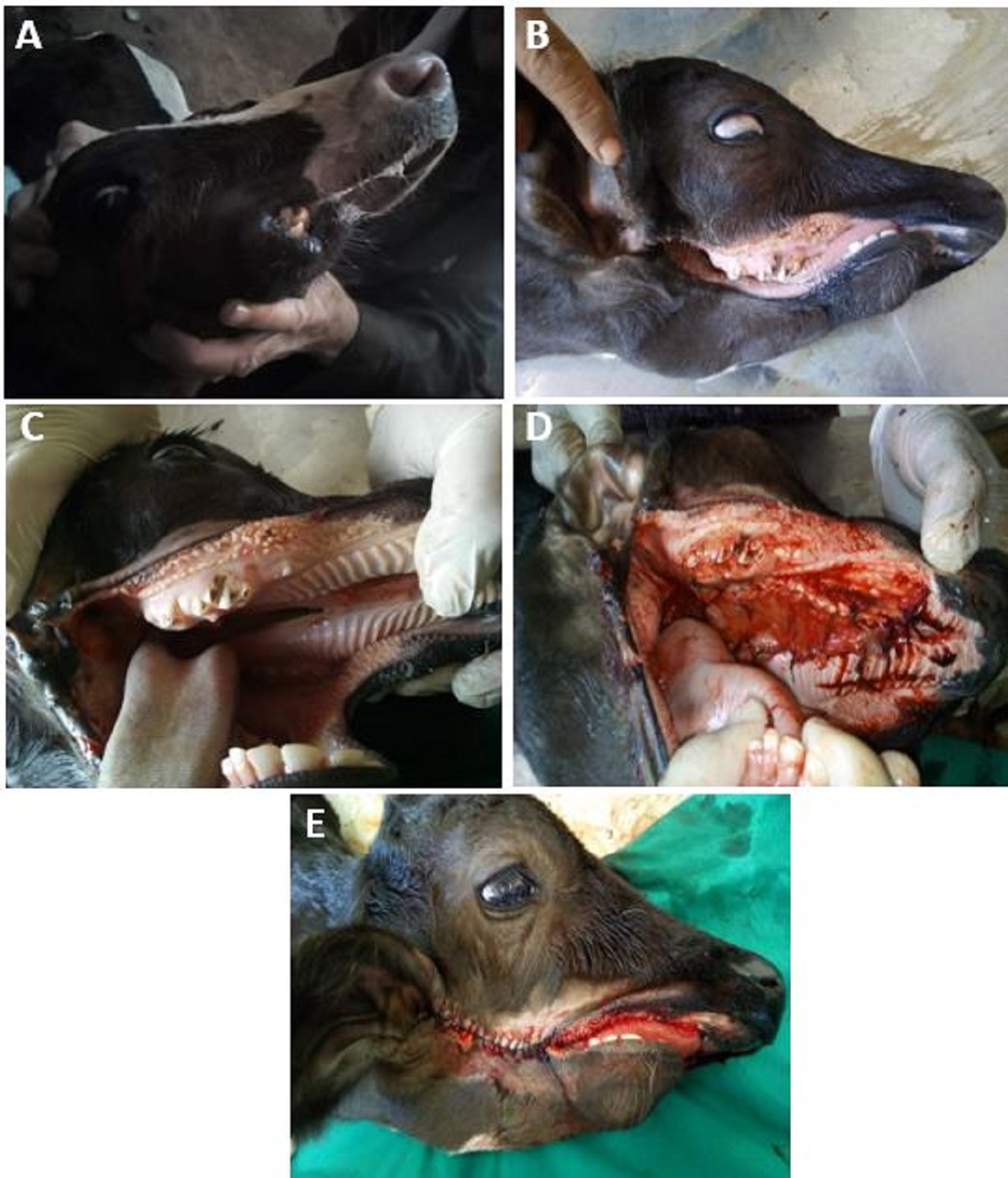
Animals and selection of cases A total of twenty-three large ruminants were selected from more than 3225 different species animals admitted to the Veterinary Teaching Hospital (VTH), Assiut University, Egypt in the duration from July 2015 to September 2019. The selection of these animals depended on the rarity of the occurrence of the included surgical disorders. The study comprised the unusual surgical conditions of the digestive system in cattle and buffaloes, these uncommon disorders were either congenital or acquired. The selected cases of this study were either rare cases or they presented with unusual signs. Species (cattle or buffaloes), breed, age, sex, chief complain, the unusual criteria, diagnosis, treatment and outcomes of all cases were recorded. The surgical disorders were diagnosed through the case history, clinical examination, exploratory puncture, radiography and ultrasonography in some cases, and the surgical findings. Follow-up information was obtained from the owners and referring veterinarians via telephone contact questionnaires. This study was approved by the Ethics Committee on Animal Experiments at the Faculty of Veterinary Medicine, Assiut University, Egypt. In addition, oral and written consents were obtained from the owners before surgical intervention. The surgical management Surgical disorders of the oral cavity (3 cases) The mandibular brachygnathism (Fig. 1A) was recorded in 2 months old mixed-breed calf. It was not treated. Twenty days old calf had not a right side commissure with right cleft extended to the right ear (Fig. 1B-D). It was treated surgically through the surgical refreshment of the edges of the congenital defect and then closed in two layers from the caudal to the cranial ends till the level opposite to the contralateral left side commissure. The inner layer included the buccal mucosa and the buccal muscles, which were sutured in a simple continuous pattern using the polyglactin 910 no. zero. The skin of the upper and lower sides was sutured in a simple continuous pattern using braided silk no. 1 (Fig. 1E). The same case suffered a severe degree of cleft palate (oro-pharyngeal fistula). It was closed by using a graft obtained from the fascia lata. The graft was anchored to the edges of the cleft palate using interrupted stitches and polyglactin 910 no. zero (Fig. 1C,D). 1ml/25 kg of penstrep (Procaine penicillin (8mg/kg)-dihydrostreptomycin (10 mg/kg); Norbrook® Laboratories Limited, Newry, United Kingdom and 2ml/45 kg of flunixin (flunixin meglumine 50 mg/1ml; Norbrook) were prescribed post-operation for 5 and 3 consecutive days respectively.
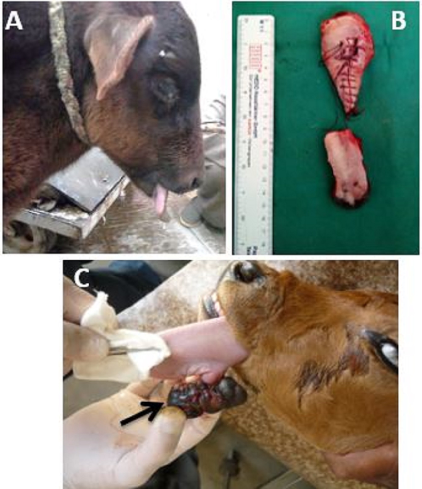
Fig. 1. (A) Brachygnathism in 2 months mix breed male calf; (B& C) Brachygnathism, wry mandible, cleft palate and opened mouth commissure in 20 days mix breed male calf. (D& E) Surgical treatment of cleft palate and opened mouth commissure. The calf suffered macroglossia (Fig. 2A) was clinically examined. The transmandibular finger test (Putter, 2011) was carried out to detect any abnormal intermandibular swelling. The macroglossia was treated by partial glossectomy (Fig. 2B) as that was described in dogs (Radlinsky and Fossum, 2019). Briefly, a tourniquet was applied around the tongue near its base. A transverse line of linear infiltration anesthesia rostral to the tourniquet was performed using 1% lignocaine HCl (Lignocaine and Adrenaline; Norbrook Laboratories Limited, Newry, Co. Down, BT35 6JP). The blood vessels on the two sides of the median line of the tongue were ligated on the ventral lingual surface using polyglactin 910 no. zero. A V shaped incision was implemented on the ventral aspect of the tongue. The acute angle of V incision was toward the pharynx and at the level of point of ligatures of blood vessels. The incision was increased in depth to reach the dorsal surface of the tongue. The piece of tongue was removed. The tips of the two arms of the remaining free portions of the tongue were apposed using one simple interrupted stitch. The tongue wound was closed dorsally and ventrally using deep continuous mattress suture pattern and polyglactin 910 no. zero. 1ml/25 kg of penstrep (Procaine penicillin (8mg/kg)-dihydrostreptomycin (10 mg/kg); Norbrook® Laboratories Limited, Newry, United Kingdom and 2ml/45 kg of flunixin (flunixin meglumine 50 mg/1ml; Norbrook) for 5 consecutive days. The tumor which was attached to the tongue (Fig. 2C) was excised surgically after the application of horizontal mattress stitches between the base of the tumor and tongue tissue using polyglactin 910 no. 1. The tumor was excised about 5 mm above the stitches. The tongue was examined carefully for any point of hemorrhage.
Fig. 2. Different affections related to the tongue. (A) Macroglossia in 2 months male calf. (B) Free portion of the tongue after 2 times glossectomies. (C) Abnormal mass (tumor) attached to the tongue of 1 month calf. Affections of the salivary ducts (2 cases) The study included a case with the dilatation (ectasia) of the parotid duct (Fig. 3A). The history, clinical examination, aseptic centesis and sialography were used for diagnosis. The latter was carried out through injection of the contrast medium (Sodium diatrizoate; Urographin 76%, SCHERING, Germany) directly into the swelling (Fig. 3B). It was managed by surgical exposure of the swelling after routine aseptic preparation. The redundant tissue was removed after its blunt dissection. An intra-oral fistula was performed according to Misk et al. (2014). The case was re-admitted after two-week post-operation with large swelling at the site of operation. Centesis was carried out under aseptic condition revealed watery whitish color fluid with flakes. The swelling was treated as abscess. It was lanced at its lower aspect. The contents were evacuated, and then the lavage of the cavity was performed using diluted povidone- iodine (5%). A drain was applied. The owner was advised for routine dressing. 1ml/25 kg of penstrep (Procaine penicillin (8mg/kg)-dihydrostreptomycin (10 mg/kg); Norbrook® Laboratories Limited, Newry, United Kingdom and 2ml/45 kg of flunixin (flunixin meglumine 50 mg/1ml; Norbrook) were prescribed post-operatively for 7 and 3 consecutive days respectively.
Fig. 3. A) Ectasia of parotid duct in 8 months female calf extended from the base of the ear distally and caudally to the retromandibular space. B) Sialography of the same case showed a large round radiopaque structure (seat of ectasia) followed with dilated parotid duct (arrows). A three-years old native breed pregnant (5 months) cow was admitted with supraorbital swelling and small opening discharging watery colorless secretion (Fig. 4A). The ultrasonography was used for examination of the swelling (5MHz convex transducer) (Fig. 4B). The case was diagnosed as sialocele, which was associated with salivary fistula. The animal was treated surgically through the opening of the swelling at the most ventral aspect and application of a drain after the evacuation of the contents. The salivary fistula was treated through the surgical ligation of the salivary duct using polyglactin 910 no. 2. A fusiform incision then excision was performed in the skin around the fistulus opening. A metal surgical probe was inserted in the salivary duct through the outer opening of the fistula. The salivary duct was ligated twice using polyglactin 910 no. 2. The subcutaneous tissue and the skin were sutured using interrupted stitches using polyglactin 910 no. 0 and braided silk no. 2 respectively. 1ml/25 kg of penstrep (Procaine penicillin (8mg/kg)-dihydrostreptomycin (10 mg/kg); Norbrook® Laboratories Limited, Newry, United Kingdom and 2ml/45 kg of flunixin (flunixin meglumine 50 mg/1ml; Norbrook) were prescribed for 5 days post-operation.
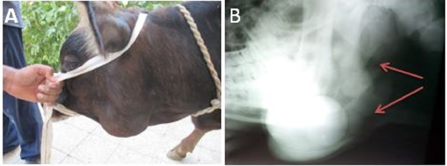
Fig. 4. A) Round swelling at the base of the ear (white arrow), dorsal and lateral to the eye (sialocele); red arrow indicates the seat of salivary fistula (wet and stained hair). B) Ultrasonogram of the sialocele showed anechoic cavity (A) enclosed within a hypoechoic wall (white arrows) using 5 MHz convex transducer. The peri and para-esophageal masses (3 cases) Three cases with peri and para-esophageal swellings were managed surgically. The para-esophageal septic hematoma in bull (Fig. 5) was opened at the most caudoventral aspect and its contents were evacuated carefully. The diluted povidone- iodine (5%) was used for lavage. A drain was applied and the owner was advised to change it as was prescribed by the surgeon. The peri-esophageal abscess (Fig. 6) in the pregnant Frisian cow was discovered incidentally after the surgical ventrolateral exposure of the esophagus under the effect of local infiltration of anesthesia using lignocaine HCl 2%. The ventrolateral exposure between the sternocephalic muscle and the trachea was performed according to Ducharme et al. (2017a). The gentle grasping of the esophagus after the blunt dissection of the muscles and fascia led to the opening of peri-esophageal mass incidentally and escaping of pus (yellowish-brown in color). The abscess was completely evacuated and the whole wound was flushed using diluted povidone- iodine solution 3%. Simple interrupted stitches and polyglactin 910 no. 2 were used for closure of the inner layers of the wound, while the skin was sutured using braided silk no. 2, leaving about 3 cm at the most lower aspect of the wound was left without suturing for drainage. 1ml/25 kg of penstrep (Procaine penicillin (8mg/kg)-dihydrostreptomycin (10 mg/kg); Norbrook® Laboratories Limited, Newry, United Kingdom and 2ml/45 kg of flunixin (flunixin meglumine 50 mg/1ml; Norbrook) were used post-operation for 5 days.
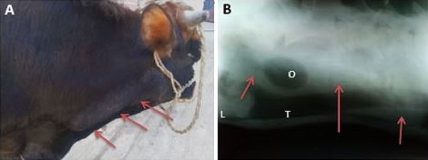
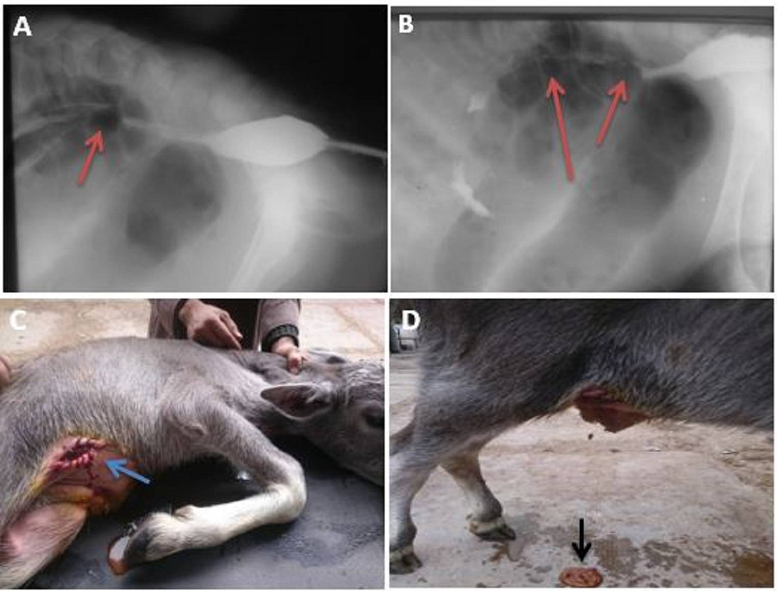
Fig. 5. A) A longitudinal uniform swelling (para-esophageal hematoma) on the right side of the neck of 1 year bull, at the level of jugular furrow extended from the mandible to about 40 cm of the neck (arrows). B) The swelling in Fig. A appeared as a radiopaque heterogenous swelling (arrows) causing ventral deviation of the trachea (T) just caudal to the larynx (L). An ovoid radiolucent structure (O) was apparent at the cranial aspect of the radiopaque swelling.
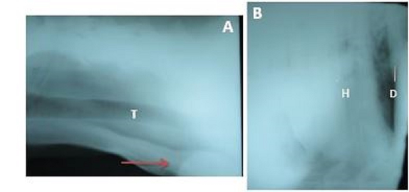
Fig. 6. A) Peri-esophageal abscess (arrow) in cow at the thoracic inlet causing dorsal deviation of the trachea (T). B) The lateral radiograph of the same animal showed the increase of the radiodensity of the thorax except the triangular area between the heart (H) and diaphragm (D) due to aspiration pneumonia. The large cervical swelling in 7 months calf (Fig. 7) was surgically managed under the effect of local infiltration anesthesia using lignocaine HCl 1% after aseptic preparation. About 9 cm skin and s/c incision over the swelling was performed followed by sharp and blunt dissection of the underlying tissue. The capsule of the cervical swelling was exposed and opened. The cervical mass was removed surgically in pieces till its complete excision without the removal of its capsule, which was firmly attached to the underlying tissue. The wound was closed in layers using continuous suture pattern and polyglactin 910 no. 2 except its lower aspect for drainage. 1ml/25 kg of penstrep (Procaine penicillin (8mg/kg)-dihydrostreptomycin (10 mg/kg); Norbrook® Laboratories Limited, Newry, United Kingdom was prescribed for 10 days post-operation for management of the generalized peritonitis.
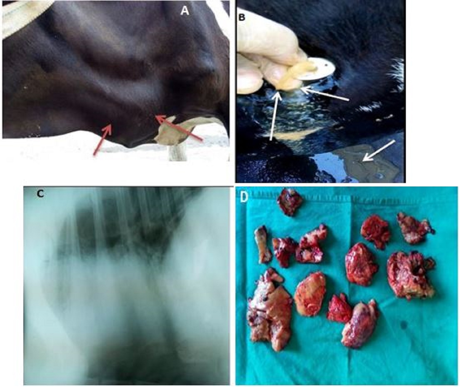
Fig. 7. A) Large cervical mass (arrows) at the lower aspect of left side of the neck (para-esophageal mass). B) Trocarization of the abdomen at the dorsal aspect of paralumbar fossa resulted in yellow color fluid (arrows). C) lateral radiograph of the thorax and cranial abdomen of 6-month calf suffered para-esophageal swelling showed disappearance of cardiac outline and increase the opacity of the ventral cranial and caudal lung lobes causing border effacement with the abdomen. D) The mass in pieces after its surgical removal. Diaphragmatic hernia in 15 days calf (1 case) A diaphragmatic hernia was diagnosed to occur unusually in a 15-days-old Frisian calf during survey radiography of the thorax. The calf was admitted with a severe degree of lameness at the right forelimb. Two lateral views were obtained (Fig. 8). It was not subjected to any treatment.
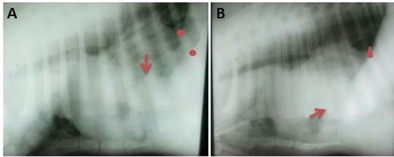
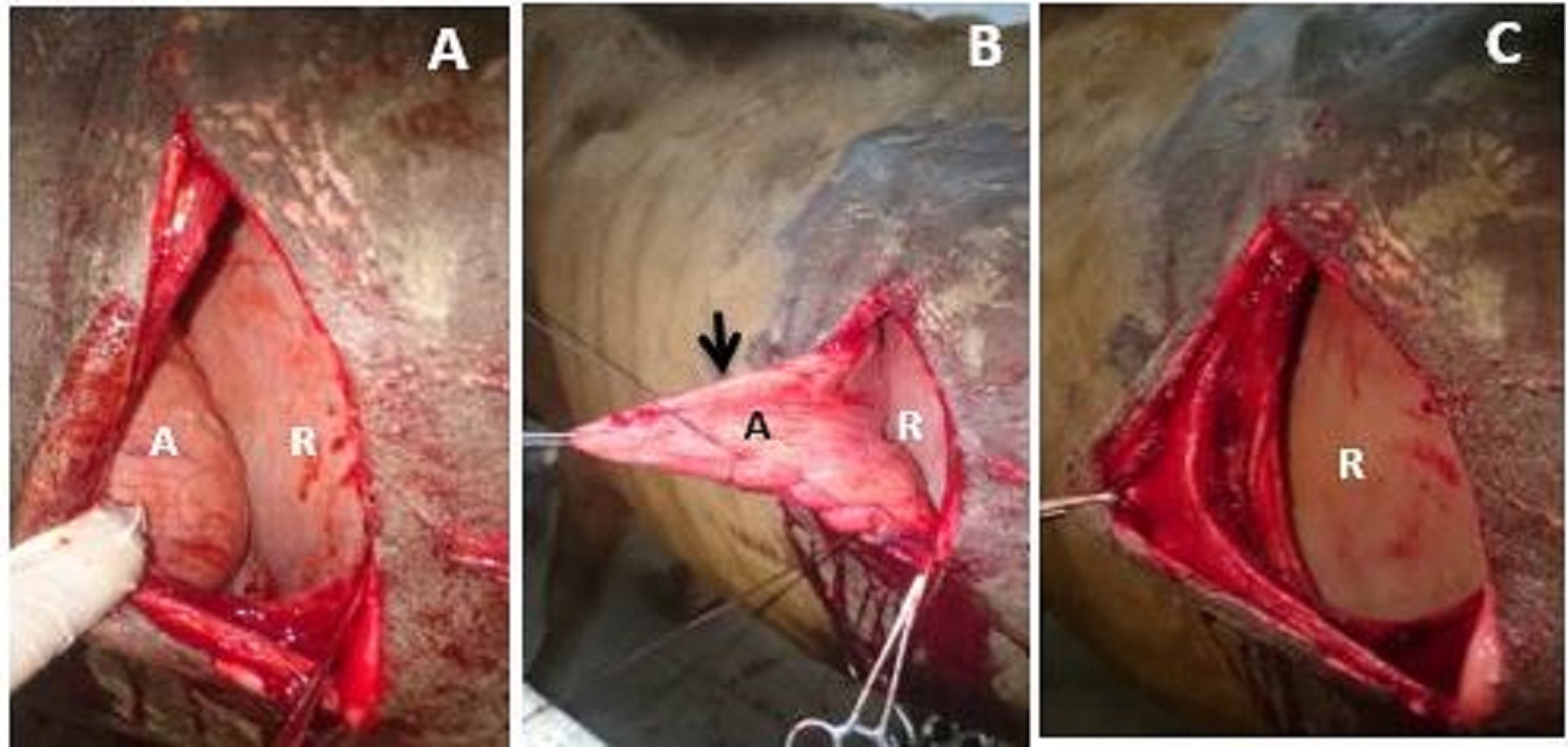
Fig. 8. Right lateral radiograph (A), and left lateral radiograph (B) in 15 days male cow-calf showed diaphragmatic hernia (arrow) and increase of the radiographic opacity in thorax. two small circles in Fig. (A) refer to two crura of diaphragm and small red triangle in Fig. (B) indicates the diaphragm as one line. Left displacement of abomasum (6 cases) Left displacement of abomasum (LDA) was diagnosed incidentally in six cows, which were decided to have laparorumenotomy. The animals underwent to the clinical (6 animals) and radiographic examination (4 animals). All the animals were prepared for surgery depending upon the consent obtained from the owners. The technique of left paralumbar fossa abomasopexy (Utrecht method) was carried out for the treatment of LDA according to Weaver et al. (2005) under the effect of local infiltration anesthesia (Fig. 9). The Weingart’s technique for rumenotomy was performed in all animals after abomasopexy according to Weaver et al. (2005) and Chaudhary (2016). The fluid therapy (20 ml/kg of normal saline 0.9%; Egypt Otsuka pharmaceutical Co. S.A.E. 10th of Ramadan City, Cairo, Egypt), 2ml/45 kg of flunixin (flunixin meglumine 50 mg/1ml; Norbrook) and 1ml/25 kg of penstrep (Procaine penicillin (8mg/kg)-dihydrostreptomycin (10 mg/kg); Norbrook® Laboratories Limited, Newry, United Kingdom) were prescribed for 2, 3 and 5 successive days post-operation respectively.
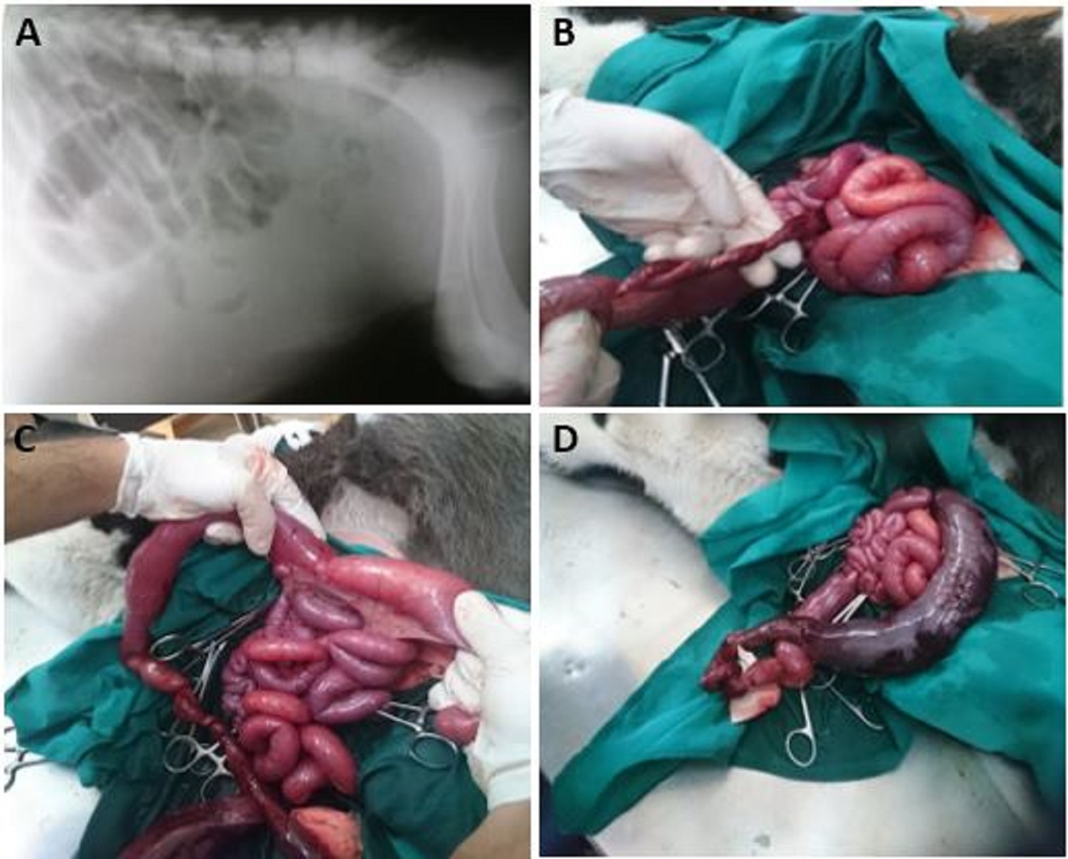
Fig. 9. Abomasal displacement in non-pregnant emaciated 5 years cow. A) It shows the displaced abomasum (A) lateral to rumen (R). B) It shows the deflation and suturing of the abomasum (arrow). C) It shows the disappearance of the displaced abomasum after its reduction and fixation into normal position (abomasopexy). Congenital intestinal volvulus (1 case) A newborn mixed-breed calf presented with atresia ani (Fig. 10). The clinical examination revealed that the animal had atresia recti. The mid abdominal celiotomy was carried out under the effect of the linear infiltration anesthesia using ½ % lignocaine HCl. Other intestinal lesions including intestinal volvulus were noted. The animal subjected to intestinal resection and creation of colonic fistula (colostomy) according to Weaver el al. 2005 and Abdel-Hakiem and Aref (2012). Congenital stenosis of the caudal segment of the colon in a buffalo calf.
Fig. 10. Radiograph of abdomen in one- day cow calf showed absence of intestinal segments (notice the gas within the intestine) (A). Twisting of segment of large intestine (B). Cecum and anterior segment of colon between two hands of surgeon (C). A dark colored blind end caudal segment of colon (may be dead) (D). The case was diagnosed through the clinical examination and the use of rectal cleansing enema (warm water and soap), and barium enema (Barium solution; 20% w/v; in a dose 10ml/kg) (Fig. 11A, B). The typhlostomy (Fig. 11C, D) was carried out according to Abdel-Hakiem and Aref (2012).
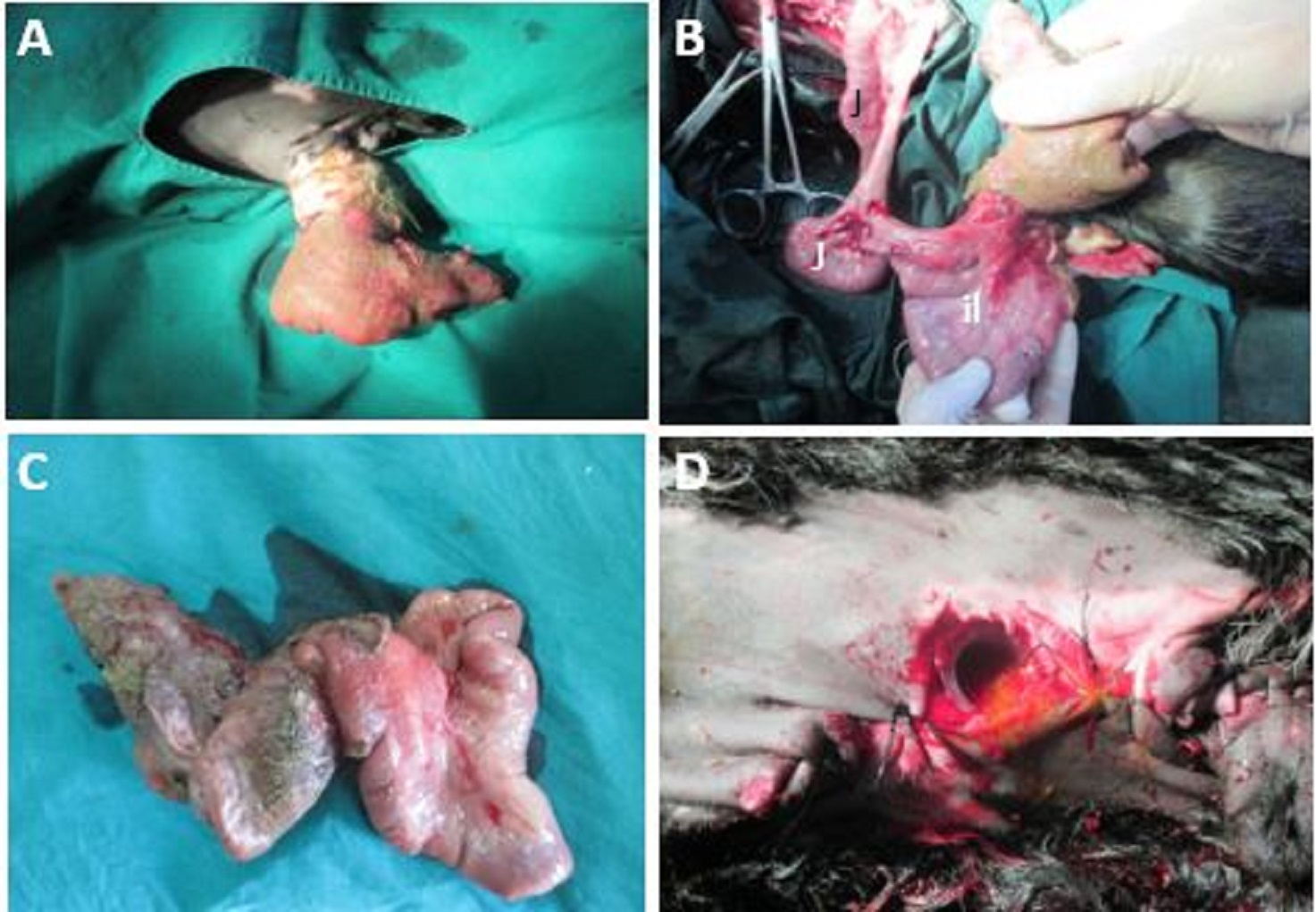
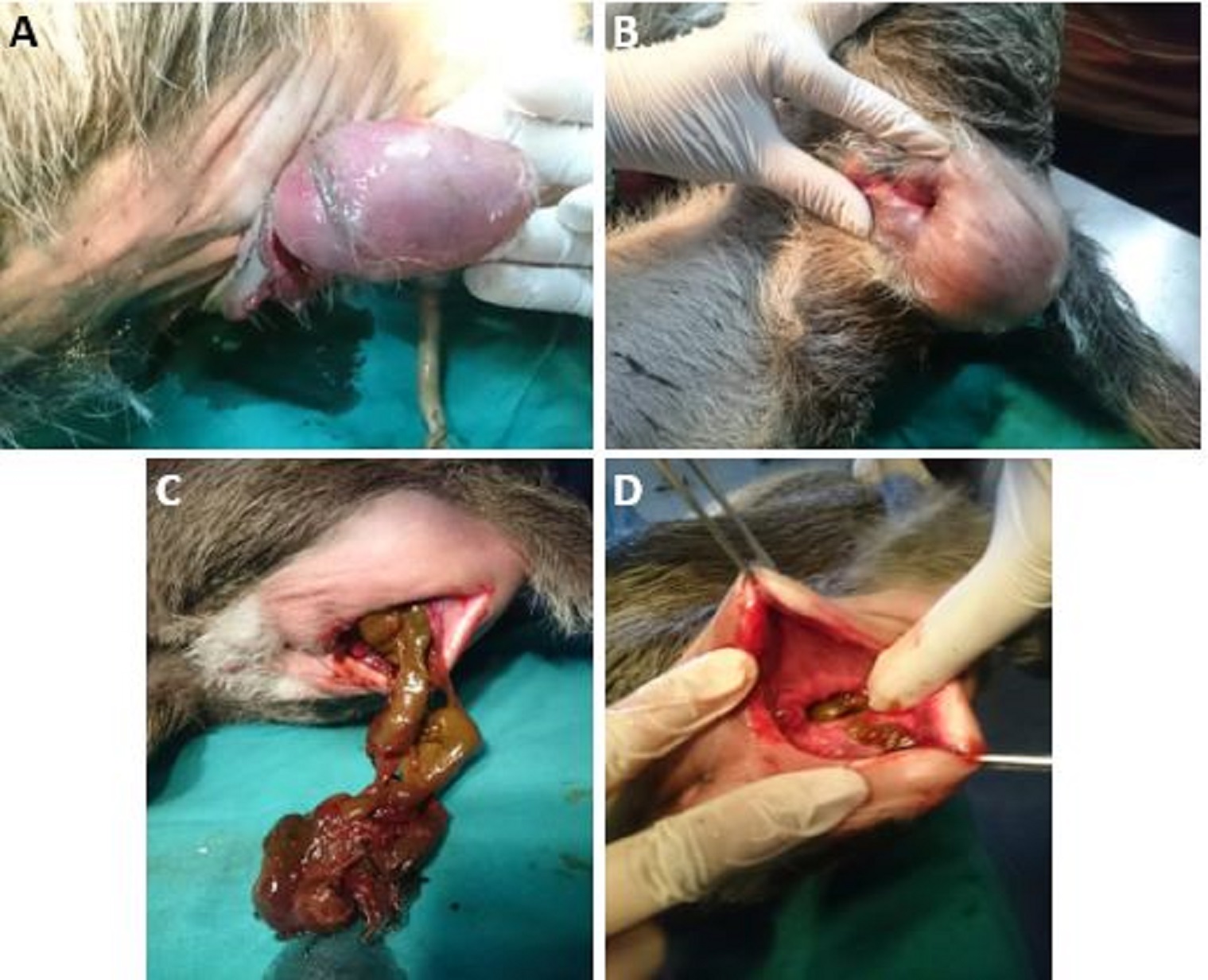
Fig. 11. Rectal barium enema showed the area of filling defects (arrows) which indicate the stenotic lumen of descending colon (A,B). Surgical created cecal fistula (typhlostomy; arrow) at the lower right flank in one-day buffalo calf (C). The animal after standing from operation and meconium (arrow) discharged from the cecal fistula (D). Umbilical eventration (5 cases) Five new-born calves had umbilical eventration (prolapse of internal organs especially the intestine) were included in the present study (Figs. 12,13,14). Four animals were subjected to surgical intervention depending on the clinical examinations. The prolapsed tissues either covered (3 cases) or not (2 cases) were cleaned and washed gently using warm distilled water. The covered membrane was peeled off in case of covered prolapsed tissues, and the contents were examined carefully. The rent of the abdominal wall was enlarged under aseptic condition and local infiltration anesthesia using lignocaine HCl ½ %. The prolapsed organs were reduced to its normal position (2 cases; liver prolapse and one case rectal diverticulum) and the other two cases were subjected to intestinal resection (edematous, highly contaminated intestinal segments and darker in color relative to the intestinal segments within the abdomen) and creation of intestinal fistula. The abdominal wall was closed in the usual manner using continuous simple pattern and polyglactin 910 no. zero. 1ml/25 kg of penstrep (Procaine penicillin& dihydrostreptomycin; Norbrook® Laboratories Limited, Newry, United Kingdom) was prescribed 5 successive days post-operation.
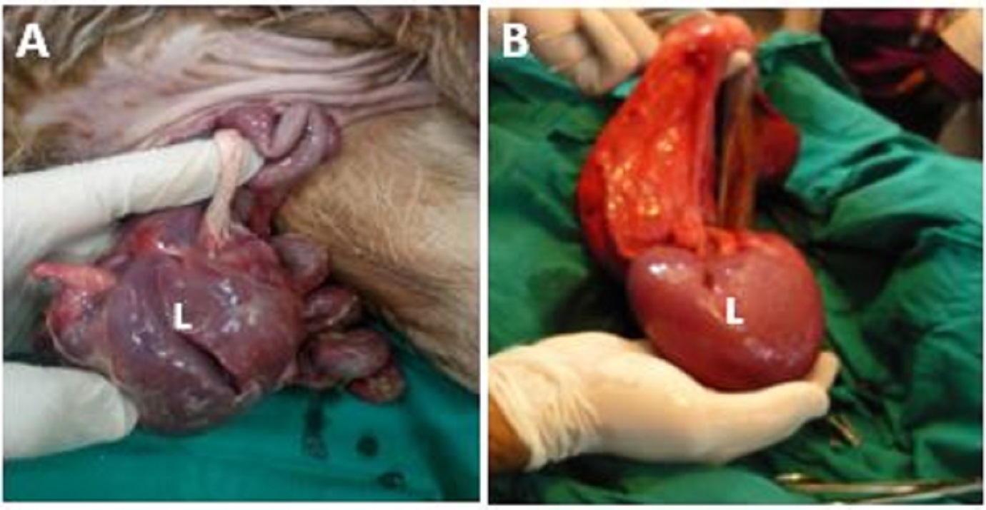
Fig. 12. Two different cases with congenital liver (L) prolapse.
Fig. 13. A) Umbilical eventration in newborn calf. B) The midabdominal laparotomy revealed the caudal end part of intestine were the jejunum (J) and ileum (il). C) The resected prolapsed part (ileum and devitalized cecum). D) Creation of intestinal fistula for discharge.
Fig. 14. Covered umbilical eventration (alatnoic sac enclosed intestine) in one-day buffalo calf (A). Atresia ani associated with outpouch (diverticulum) of rectum (B). Surgical incision of outpouch and fecal escaped out (C). Excess skin (holded by allis forceps) was removed and remained skin was sutured to the rectal wall circumferncially (D). |
|
|
|
Results |
|
The study included 23 animals with different surgical affections of the digestive system. They were 22 cattle and 1 buffalo species. The total number of male animals was 17 (16 cattle and 1 buffalo species), while the female animals were 6 cows. The ages of animals ranged from a few hours (newly born) to 5 years old. Their weight ranged from about 40 to 350 kilograms. Some of the animals (n= 7) suffered a single type of surgical affection, while the others (n= 16) had multiple lesions. The congenital lesions were recorded in 12 animals, while 10 animals suffered from acquired disorders. One case could not be determined either a congenital or acquired. Twenty animals were treated surgically. All the treated animals were followed up. Eleven animals were improved. The other nine animals either died at different times post-operation (n.=8), or was not improved (n.= 1). Left displacement of the abomasum represented the digestive lesion with a high successful rate of recovery (5/6; 83.3%). Cases of intestinal agenesis and umbilical eventration were the digestive lesions with a low rate of improvement and recovery (6/7 died; 85.7%). The aseptic centesis (exploratory puncture) was used for 4 animals (ectasia of parotid duct (1), sialocele (1), para-esophageal hematoma and peri-esophageal mass (2)). It supported the final diagnosis in 2 cases (ectasia and para-esophageal hematoma). The mandibular brachygnathism was congenital affection in a mixed breed male calf. It was calved from its dam after the full term pregnancy, which resulted from normal mating. It was the 2nd parturition that occurred normally without any external interference. The animal showed disability for drinking and suckling. The examination of the calf at two months post-parturition showed its awareness and normal activity but its body weight was decreasing compared to its peers. The incisors were irregular in position and overcrowded. There was decrease number of the lower cheek teeth (molar and premolar). The mandible was shorter than the maxilla about 11 cm (Fig. 1). It was slightly deviated to right (wry mandible). There were no other defects associating the mandibular brachygnathia. The calf’s health condition was progressively deteriorated. When the calf completed its fourth month, it was recumbent and unable to stand by himself. The calling of the owner about 1 year of the animal’s admission displayed that the mother gave a normal third birth. A 20-days mixed breed male calf was referred with a defect of the right cheek wall, which extended from the point of absent mouth commissure cranially to the level of the ipsilateral ear canal caudally. The calf had other associated anomalies such as right deviation of the mandible and severe degree of cleft palate (oronasal fistula) (Fig. 1B-E). The chief complaint was the decreasing ability of the animal for drinking and suckling. The animal had no capability to keep the milk in its oral cavity during external feeding. The animal was alert and had normal condition and physical parameters. The lateral opened cheek and the cleft palate were closed surgically. Intra-operative hemorrhage was extensive especially during the reconstruction of the cleft palate after the incisions of the hard and soft palates on the both sides of the cleft (fistula). The post-operative following up recorded the death of the animal at the 4th day of operation. The calf with macroglossia (long tongue) was alert but dehydrated. The heart rate was regular, strong and rapid (100 beats/minute).The owner reported decrease of drinking and suckling behavior. The tongue was protruded out of the mouth cavity most of times. The tip of the tongue protruded about 7 cm past the mandibular incisors passively without its traction (Fig. 2A, B). The tongue was protruded centrally not to any side (Fig. 2A). The animal suffered continuous ptyalism. It was able to curl and retract its tongue to its normal intraoral position. The thorough examination of the oral cavity, tongue and teeth revealed no other associated lesions. The transmandibular finger test detected no ventral bulging at the intermandibular space and the floor of the mouth. Post-operation, the partial resected tongue was within the oral cavity. There was no hemorrhage after amputation. The animal was followed up post-operatively. The owner reported there was no obvious improvement in the drinking and suckling behavior of the calf. Eventually, it survived for twenty days after surgery then died. The mass attached to the tongue in the Brown swiss male calf was congenital in origin. It originated at the left border of the tongue about 4 cm from its tip. It had a well-demarcated neck. The mass was fissured or lobulated externally, which looked like the kidney of cattle (Fig. 2C). The length of the mass extended from the left mouth commissure to the calf’s chin. The tongue at the point of mass attachment was apparent even with the closed mouth. The mass was resected easily with control of hemorrhage. It weighted about 150 grams. The case improved and the suckling behavior became normal. The radiography and contrast radiography were used for 11 animals [ectasia of the parotid duct (1), Para and peri-esophageal masses (3), diaphragmatic hernia (1), LDA (4), intestinal volvulus (1), and colonic stenosis (1)]. It determined and confirmed the final diagnosis in 5 out of 11 animals. They included ectasia of the parotid duct (1), Para esophageal masses (2), diaphragmatic hernia (1) and colonic stenosis in buffalo calf (1). The ectasia of parotid duct was recorded in 7 months female Brown Swiss calf. It appeared as fluctuating large elongated swelling. It was on the left side. The swelling was mostly retro-mandibular in position (Fig. 3A). The centesis of the swellings revealed colorless watery viscous fluid. The sialography revealed that the dimensions of the dilated parotid duct (diverticulum) were 9 x 11.5 cm (Fig. 3B). The heights of the parotid duct caudal to the point of ectasia were 3.8 (Fig. 3B). Salivary sialocele and fistula were diagnosed in a 3 years-old cow. The swelling was rostral to the base of the ear. The swelling was well-demarcated, circumscribed, extended from the base of the left ear to the lateral canthus of the left eye and lateral to the temporal line of the frontal bone (Fig. 4A). It was fluctuating even though the tension of skin overlying it. The aseptic centesis of the swelling resulted in a colorless watery viscous fluid that resembled the saliva. The ultrasonographic examination revealed anechoic homogenous contents encased in a hypo-echogenic wall with no apparent acoustic enhancement (Fig. 4B). The salivary fistula was on the same side of the swelling, about 3- The para-esophageal septic hematoma appeared as elongated uniform swelling on the right side of the neck. It extended from the retropharyngeal space to about 40 cm caudally (Fig. 5A). It was on the level of jugular vein. It looked on the lateral radiograph as long heterogeneous radiopaque mass (38x 6.5 cm) at the level of the esophagus, dorsal to trachea and extended from the level of cervical vertebra no. 1 to the cervical vertebra no. 5. A radiolucent oval structure (5x7.5 cm) within the heterogeneous mass at the level of cervical vertebra no. 2 appeared on the radiograph. There was a ventral deviation of the trachea at the level of radiolucent oval structure, but a slight dorsal deviation of the trachea was noticed caudally (Fig. 5B). The cow with the peri-esophageal abscess had the signs of esophageal obstruction such as pyalism, tympany and difficulty to introduce the gastric tube. There was a gradual decrease of feed intake, which started 1 week before admission and completely stopped at the day of admission. The typical signs of esophageal obstruction appeared few hours before coming to the hospital. The radiographic examination revealed a round radio-opaque structure (9x7 cm) ventral to the trachea, just in front of the thoracic inlet, opposite to the cervical vertebra no. 6. It produced a slight dorsal deviation of the trachea (Fig. 6A). The radiographic examination of the thorax showed an increase density especially the cranio-ventral aspect of the lungs (Fig. 6B). During the surgical exposure of the esophagus yellowish-brown color pus with offensive odor was escaped through the surgical wound. The animal was followed up post-operatively, and the owner reported the recovery of the animal within three days after operation. The peri-esophageal mass in the six-months calf was clear on the left side of the neck. It was circumscribed in its appearance. It extended from the mid-neck region to just in front of the point of shoulder. Its dorsal wall was at the level of jugular vein (Fig. 7A). The animal had signs of esophageal obstruction such as tympany, decrease to loss of feed intake. The animal’s condition was bad and its physical signs were (T= 39.5°C, HR= 85 bpm, RR= 26 rpm). The calf was dehydrated (6%) (Skin tent duration was 5 seconds; the eyeball was mildly sunken). The trocarization at paralumbar fossa resulted in copious amount of yellow color watery fluid with offensive odor and small flakes (Fig. 7B). Radiographically, the mass appeared as a radio-opaque homogenous structure causing dorsal deviation of the trachea. The lateral radiograph of the thorax and cranial abdomen showed increase opacity of the thorax especially at the ventral aspect of the cranial and caudal lung lobes. The cardiac outline was not clear. Border effacement is detected at the area enclosed the caudal border of the heart, diaphragm and the cranial abdomen (Fig. 7C). The mass was surgically excised completely but in pieces leaving its capsule (Fig. 7D). The animal died 24 h post-operation. The nature (hematoma, fibrous tissue) of the para/peri-esophageal masses could not be determined by radiography, but the presence of radiolucent oval structure in case of para-esophageal hematoma referred to infection that may be gas-forming microorganisms. The calf with diaphragmatic hernia (DH) was alert, and its health condition was normal. The temperature, heart rate, and respiratory rate were 37.5 °C, 90 bpm and 22 rpm respectively. The clinical examination of the right fore-limb revealed abnormal passive movements of the carpal joint. A deformity of the proximal aspect of the cannon bone (3rd metacarpal bone) was detected. Radiographic examination showed an old healed fracture at the proximal third of the right metacarpal bone. The appearance of the DH on the right and left lateral radiographs was nearly the same. Discontinuation of the diaphragm was noticed on the radiograph. There was a circumscribed swelling (12x 5.5 cm) in front of the diaphragm which merges with the caudal border of the heart. The distance of this structure (distal aspect) from the sternum was almost the same (7.5 cm) on the two views. The outline of the heart was deformed and could not be determined exactly. The density of the thoracic cavity was generally high and appears more radio-opaque than normal. The lumen of the caudal trachea looked more radio-opaque on the right lateral views (Fig. 8). All cases of LDA admitted with chronic decrease of feed intake and mild degree of left flank tympany which were not responding to medical treatment. The owners noticed low rumination and scanty moist feces of their animals. The auscultation of the rumen showed either no audible ruminal cycles or low pitched cycles as if heard from far distance. All the animals suffered 6 % dehydration, and high pitched tachycardia (90-95 bpm). Also, a well demarcated round structure at the dorsal left flank was felt percutaneous in three cases. Five out of six surgical treated animals recovered within the first week of operation. The remained animal died within 2 days of surgery. The left displacement of the abomasum (LDA= 6/24 or 25%) and the umbilical eventration (5/24 or 20.8%) represented the high percentage of the surgical affections in this study. All cases of LDA were acquired, while the five cases of umbilical eventration were congenital in origin, and associated with other lesions. Two cases had liver prolapse, two had complete agenesis (absence) of large intestine and one case suffered atresia ani and rectal diverticulum. The numbers of surgical affections out and within the abdominal cavity were 11 (45.83%) and 13 (54.17%) respectively. The calf had congenital intestinal volvulus and subjected to intestinal resection and colostomy died within few h post-operation. The animal suffered atresi ani et recti and discoloration (dark red color) of the blind end caudal colon (Fig. 10). The buffalo calf suffered severe stenosis of the caudal segment of the descending colon died on the day after the day of operation. The diagnosis of this case was through the rectal enema, contrast radiography and exploratory laparotomy. The calf had a patent anal opening and rectum. The connection between the rectum and the last portion of the descending colon was cord like and impervious except for traces of barium solution. This portion appeared radiographically as filling defect opposite to the intervertebral disc between the fourth and fifth lumbar vertebrae (Fig. 11 A, B). The typhlostomy discharged meconium immediate post completion of the surgery (Fig.s 11 C, D). The umbilical eventration was recorded in five cases. The liver prolapse (Fig. 12) was observed in two calves (male and female). The follow up of the treated case revealed that the animal recovered and became in normal condition. The clinical parameters of the other case was abnormal (T= below 35°C, RR= 7 rpm; slow deep). The animal died within 1 hour after its admission to the hospital. The two cases of complete agenesis of large intestine (Fig. 13) died on the same day of operation. The calf had absence of the anal opening, diverticulum of the anal canal, and covered umbilical eventration (Fig. 14) became in normal condition after operation. |
|
|
|
Discussion |
|
The present work included large ruminants that had unusual surgical disorders of their digestive system or which were admitted with unusual presentation with special references to their diagnosis, treatment and outcomes. The recorded results of this study revealed that 66.67% of the animals were male. These results were consistent with the results of previous studies (Magda and Youssef, 2007; Abdel-Hakiem and Aref, 2012; Patil et al., 2016; Abdelhakiem and Elrashidy, 2017). This may be due to the increase number of young animals included in the study relative to the mature animals. Sixteen out of twenty-three animals in this study have the age of eight months or lower. The number of cattle with surgical disorders in this study was higher than the number of buffaloes. The mandibular brachygnathism (brachygnathia inferior, parrot or overshot mouth, micrognathia) is the shortage of the mandible relative to the maxilla. It was reported that this condition may be congenital or acquired in origin (Dotzel and Rawlinson, 2017). In fact, this anomaly is considered one of the multiples that occur in farm animals. So, the real cause behind this anomaly could not be elucidated. The disturbance of development of different organs and systems may lead to single or multiple defects (Vegad, 2009). Some of the infectious agents (Orthobunyavirus Schmallenberg virus and the virus of Bovine viral diarrhea/mucosal disease) were incriminated in the occurrence of inferior brachygnathism (Fowler, 2010; Herder et al., 2012). It was stated that the brachygnathia inferior is considered an inherited and lethal autosomal recessive disorder in sheep and calves (Shariflou et al. 2011; Uzal et al., 2016), and it could be associated with other lesions such as cerebellar hypoplasia in calves (Uzal et al., 2016), cardiomegaly and renal hypoplasia in sheep (Shariflou et al. 2013) and other defects (Fowler, 2010; Herder et al., 2012). The case of the present study was accompanied with right deviation of the short mandible, overcrowdings of the incisors and shortage of the tongue. The calf suffered brachygnathia in the present study was male. This is consistent with what was stated by Uzal et al. (2016). According to the obtained data about 1 year after animal’s admission, the dam of this calf had two normal calves before and after this affected calf. So, it was postulated that the brachygnathia inferior might be a sex linked disorder. Although several cases of overshot mouth were recorded in calves in the previous studies (Heidari et al., 1985; Griffith et al., 1987; TAŞAL and AYTEKİN, 2015; Uzal et al., 2016), the case of the present study was considered a rare recorded anomaly. This is due to the difference between the short mandible and normal maxilla was about 11 cm. It was considered a severe case of brachygnathia that was not recorded before. TAŞAL and AYTEKİN (2015) measured about 9 cm as a difference between the short mandible and normal maxilla. This malformation had a clinical impact on the animal health. The suckling behavior was abnormal. The animal was weak, lethargic progressively. At the end of the fourth month of his birth, it was recumbent and unable to stand with himself due to malnutrition (Murali et al., 2013). To the author knowledge and the available data in the previous literatures, the present study recorded for the first time a 20-days mix breed male calf with loss of right commissure and right lateral cheek wall of the mouth cavity. This anomaly had its adverse effect on the animal’s health (Murali et al., 2013). Due to the loss of normal suckling and drinking behavior, and the disability of the animal to keep the milk in the oral cavity after external feeding may be the most anxious problem facing the animal and his owner. The aim of the surgery was to improve the suckling behavior and reduce the losses of introduced milk either through the opened mouth commissure or the oronasal fistula. Uzal et al. (2016), reported that the formation of the jaws, oral cavity and the face of the animal during the gestation period needs an integration of the frontonasal, maxillary and mandibular embryonic processes. The defects of the formation or union of these processes led to the congenital anomalies of the skull. They added that the facial clefts may include the skin or the deeper structures. In the present study, the cleft which extended from the angle of the mouth to the ear canal might be resulted from the failure of the fusion of the lateral portions of the maxillary and mandibular processes. The other associated defects of this calf such as cleft palate (palatoschisis) and deviation of the mandible were reported in previous reports (Abdelhakiem and Elrashidy, 2017; Ducharme et al., 2017a). In the study that was conducted by Panter et al. (2000) on pregnant does and ewes, the exposure of these animals during their first trimester of pregnancy to Nicotiana glauca (wild tree tobacco) led to cleft palate in the newborns. Therefore, depending on the results of that study; the exposure of pregnant animals during the early gestation period to external harmful effect may hinder the normal palate closure and reduce the fetal movements which eventually resulted in cleft palate and other congenital anomalies such as flexural deformities (Panter et al., 2000). In Pyrenees Shepherd dogs, the cleft palate was suggested to be an inherited anomaly that occurs due to a monogenic autosomal recessive trait (Kemp et al., 2009). The case of macroglossia (tongue hypertrophy or hyperplasia) in this study was considered a rare case. It was recorded as a sole or a single congenital lesion in a native breed male calf. The term macroglossia refers to oversized tongue, or very long tongue (McGraw-Hill, 2002; Blood and Studdert, 2007; Putter, 2011). It could be congenital (Chibuzo, 1981; Singhal et al., 2016) or acquired disorder which occurred as a result of pathology of the tongue such as inflammation, abscessation, neoplasia or infectious diseases (Schoof, 1997; Hernandez and Negro, 1999, Von Doerenberg et al., 2008; Montinaro and Boston, 2013; Uzal et al., 2016). In the literature, it was recorded in calves with congenital hypotrichosis and myofiber hyperplasia (Double Muscling, Doppelender, Culard) (Oliver and Cartwright, 1968; Constable et al., 2017; Scott, 2018). It was associated with cleft palate and lip, loss of facial bones in dead cross breed cattle fetus (Singhal et al., 2016). Furthermore, it was documented in 5 years Cavalier King Charles Spaniel dog (Putter, 2011). In human, the macroglossia was recorded in previous studies. It was associated with other lesions such as congenital diaphragmatic hernia, acromegaly, muscle pseudohypertrophy and amyledosis. It was recorded as sex linked overgrowth syndrome (Hammersley and Moos, 1985; Quijano-Roy et al., 2002). In the present study, the macroglossia was the primary finding and was not associated with other defects. The tongue was very long and protruded out the mouth cavity. It looked like the case of glossplegia but the animal had the ability to curl and retract it within the oral cavity which indicated the intact of hypoglossal nerve (CN XII) (de Lahunta and Glass, 2009). Depending on the history and clinical signs, the calf was dehydrated. This may be due to the profuse continuous salivation and decrease of the water and milk intake. The follow-up of this case revealed that the calf survived about twenty days post-operation without significant improvement. The real cause beyond the death of the animal could not be determined. Ducharme et al. (2017a) reported that the tongue in large ruminants is so important for prehension. So, the tongue amputation in these animals after lacerations is associated with greater morbidity than in horse and small ruminants. Many surgical diseases may interfere with the prehensile ability of ruminants to transfer the food into the esophagus (Ducharme et al., 2017a). Any internal or external lesion of the tongue in large ruminant has a negative effect on the animal health. The present study recorded a mass attached to the tongue in a newly born calf. This mass had an adverse effect on the animal. It could not use the tongue properly. There were no available data about the congenital attachment of the tongue with abnormal masses in calves. Valentine and Barrell (2017) reported that the calves may be born with multiple cutaneous vascular tumors. It is well-known that the tumors of the epithelial origin are common (Dorn et al., 1968). The papilloma is one of the neoplasms of the epithelial origin (Pawaiya and Kumar, 2011). Valentine and Barrell (2017) stated that the papilloma can affect the oral mucosa of the calves especially the lips. It affects the mucosa of the alimentary tract of cattle and dogs (Newkirk et al., 2017). The papillomatosis was recorded as a congenital tumor of the tongue in pigs in china. In small animals, the lingual tumors are uncommon (Beck et al., 1986; Carpenter et al., 1993). In cattle practice and dogs, it was noted that the squamous cell carcinoma and liposarcoma are the common tumors originated from the tongue of mature or aged animals (Carpenter et al., 1993; Montinaro and Boston, 2013; Valentine and Barrell, 2017). The parotid salivary gland is one of the major salivary glands in bovine species. Its role is the secretion of saliva (Mansour et al., 2018). The anatomical position of the gland and its duct in ruminants were discussed and established before (Ashdown et al., 2010; Mansour et al., 2018). The ectasia or dilatation is one of the surgical affections of the parotid duct (Misk et al., 2014). Its occurrence is high in buffaloes than cattle (Misk et al., 1991, 2014; Semieka, 2002). In the present study, a congenital case of parotid duct ectasia was recorded. It might be postulated to a congenital cause that led to parotid duct ectasia causing weakness and relaxation of the wall of the duct which gradually enlarged with the time to form diverticulum enclosed saliva. The agenesis or atresia of the parotid duct papilla might be a main cause beyond the formation of fluid filled swelling (Ducharme et al., 2017a). It was reported that the partial or intermittent duct obstruction is necessary for development of the chronic dilatation of the duct. But the acute complete obstruction leads to temporary dilatation of the duct which rapidly resolved due to back pressure atrophy of the gland (Hoffer 1971; Deyoung et al., 1978; Slocombe, 1980). The obtained history referred to the chronic development of the swelling and its gradual enlargement. This verify that the seat of obstruction at the end of parotid duct (its papilla). The shape, size and extension of the dilated duct were exhibited on the sialography. The swelling was diffuse and large which extended from the base of the ear proximally to beyond the ventral aspect of the left ramus of the mandible distally. As well as, it extended caudally to the level of the pharynx. The swelling from outside as if had two compartments. These results somewhat differed from the results recorded before by Misk et al. (2014). They reported that the distended duct looked like the fest which formed on the course of the parotid duct (lateral cheek either right or left). This site is dorsal to what recorded in the present work. This may be attributed to the difference in species. The previous studies recorded most cases of parotid duct dilatation in buffaloes (Semieka 2002, Misk et al., 2014). Diagnosis of the ectasia of parotid duct in the present study depended on the characters and location of the swelling, exploratory puncture and sialography (Semieka 2002; Misk et al., 2014; Ducharme et al., 2017a). The swelling was free from the signs of inflammation. The exploratory puncture has an important role in the diagnosis. It revealed a colorless watery secretion with low viscosity. Misk et al., (2014) stated that the aspirated fluid in case of ectasia was straw yellow colored fluid. According to the text books of ruminant anatomy, the salivary gland could be classified into serous, mucous and mixed. Serous gland secretes a watery clear fluid, while the mucous gland secretes a mucous viscous fluid. The mixed gland combines a mix of two secretions. The parotid gland is considered serous gland (Frandson et al., 2010; Mansour et al., 2018). So, the aspirated fluid in the present work was consistent with the natural color and viscosity of the secretion of the parotid gland. Different treatments were documented for ectasia of the parotid duct including Intraoral marsupialization, the surgical reconstruction of the duct, application of polyethelene tube after reconstruction, destruction of the gland using irritant materials (Semieka 2002; Misk et al., 2014; Ducharme et al., 2017a). In the present study, the animal was treated using intraoral marsupialization. The animal re-admitted two weeks after with suppurative swelling. It was managed as abscess. The animal was followed up post-operatively with good results. Misk et al. (2014) reported the successful results of the intraoral marsupialization technique for management of the parotid duct ectasia in buffaloes. The second case of the salivary gland/duct affections was sialocele. It was recorded in a native breed cow. It was a large swelling at rostral base of the left ear and lateral to the left eye. It associated with a salivary fistula. The latter was recorded before especially in buffaloes (Misk et al., 2014). But, this form of sialocele was not recorded before in the literatures concerning the salivary gland affections especially in ruminants. The aseptic aspiration showed a colorless watery viscous secretion resembles the saliva. This swelling might result from either damage or rupture of the parotid gland or its duct at the duct-gland junction. This rupture led to leakage of saliva and formation of mucocele or sialocele at this region. The rupture of the gland or duct may be due to violent trauma. The presence of salivary fistula accompanied the abnormal swelling may overweigh the trauma as a real cause. Misk et al. (2014) attributed the main cause beyond the salivary fistula in buffaloes to the severe or violent trauma. Moreover, the anatomical positions of the parotid gland and its duct in large ruminants make them liable for injury due to external trauma. The present study included three cases of para and peri-esophageal masses. Two cases were male animals while the third one was Frisian cow at a late stage of pregnancy. The clinical signs in two cases were consistent with the signs of esophageal obstruction. A moderated degree of tympany (Haskell, 2008), regurgitation of food not water and difficulty in insertion of the stomach tube were noted. It was reported that the causes beyond the compression of the extrathoracic trachea may lead to compression of the esophagus causing the signs of esophageal obstruction (Braun et al., 2014; Nichols, 2017). The clinical signs and radiography were important in the diagnosis. Two cases (2 male animals) admitted with external obvious swellings on the neck. One swelling was diagnosed as hematoma, and the other was encapsulated large fibrous mass. The first case was associated with dysphagia while the latter was associated with signs of esophageal obstruction. A late stage pregnant cow was diagnosed with peri-esophageal abscess during the surgical exposure of the esophagus. This cow had signs of esophageal obstruction. According to the obtained history from the owner, cervical mass in the calf developed and enlarged progressively. Ducharme et al. (2017a) reported a case of encapsulated fibroma in young cattle that developed progressively but in a different position. The mass was recorded at the right retropharyngeal space. At the radiographic examination of the present cases, there was a dorsal deviation of the trachea in two cases. While the third case the mass did not affect the trachea. According to these radiographic findings the first two cases suffered peri-esophageal masses and the last one was diagnosed as para-esophageal mass (hematoma). These masses exerted its impact on the esophagus not the trachea which may explain the signs (dysphagia, regurgitation, tympany) associating the admitted animals. The results of this study were in accordance with the results that were suggested by Ogilvie (1998) and Rings (2009). The latter stated that the signs of esophageal obstruction may result from compression of the esophagus externally due to abscesses along the neck, especially at the thoracic inlet, or neoplasms such as lymphosarcoma (thymic and mediastinal lymph node). The peri-esophageal abscess in the present work was near to the thoracic inlet (Rings 2009; Braun et al., 2014). Two cases improved after the surgical treatment, while the third case (peri-esophageal fibrous tissue mass) died few hours post-operation. This may be due to the generalized peritonitis associated this case. As well as, the aspiration pneumonia that was confirmed on the radiographs (Cable et al., 1998; Pasquini and Pasquini, 2010). The aspirated large amount of fetid odour and whitish-green color pus from the left flank region, and generalized poor condition were consistent with signs of generalized peritonitis (Pasquini and Pasquini, 2010). The generalized peritonitis might result from the septic technique for evacuation of the gases from tympanic rumen. The ruminant contents were leaked through the ruminal wall after its trocharization. The diaphragmatic rupture or rent is one of the common lesions of the diaphragm in animals. The diaphragmatic hernia was recorded in cattle and buffaloes and the latter were reported to be more affected (Deshpande et al., 1982; Misk and Semieka, 2001; Ducharme et al., 2017b). The diaphragmatic hernia (DH) may be congenital, traumatic or hiatal in origin (Ducharme et al., 2017b). The mature cattle commonly affected by DH than young or immature animals (Misk and Semieka, 2001; Ducharme et al., 2017b). There were few cases of DH, which were recorded in newly born calves (Bellavance et al., 2010; Hicks and Britton, 2013). In the present study, the admitted animal was 15-day-old Frisian male calf. He presented with right forelimb lameness, which resulted from metacarpal fracture as confirmed by the radiograph. The DH was found out incidentally. So, the actual origin behind the DH could not be determined. It could be congenital (15-day-old calf at the day of admission), or traumatic especially it was concurrent with metacarpal fracture. It is worthy to mention that the calf had no any abnormal signs other than the right forelimb lameness. The data obtained from the owner about 100 days from the date of admission revealed that the animal survived normally without any obvious abnormal respiratory or cardiovascular signs. On the contrary, the previous studies reported the presence of respiratory (distress) and gastrointestinal signs in animals with congenital DH (Bellavance et al., 2010; Hicks and Britton, 2013). No one can deny the important role of radiography in detection and diagnosis of the DH (Misk and Semieka 2001; Bellavance et al., 2010). In the present case there was no difference in the displaying of DH on the right and left lateral views. This might indicate that the herniation was at the center of the copula of the diaphragm. In this study, six cases of left displacement of abomasum (LDA) were recorded. Three animals were males and their ages were 6, 7 and 14 months. Their breeds were mixed breed. The other three cases were cows. Their ages were around five years. One of them was native breed, and other two cases were of mixed breed. The LDA was recorded in previous studies and documented in the textbooks of large ruminant medicine. The point of difference in this study was the clinical presentations of the animals. All the animals shared the clinical signs including chronic decrease of feed intake and scanty feces. Also, the three cows and two male calves had a low body score which did not exceed number two according to Ferguson et al. (1994). The milk yield was very low in the cows, though the previous researchers reported that the LDA is a disease of cows with high milk production (Van Winden and Kuiper, 2003). In the present study one cow was pregnant (40 days), and two cows were non-pregnant. The time elapsed since last parturition was more than 6 months. Coppock (1974) recorded that the abomasal displacement occurs in dairy cows with ages ranged from 3-10 years and in the first two- four weeks post-parturition. It was reported that LDA is associated with other diseases such as ketosis, metritis, retained placenta, mastitis, hypocalcemia and Vitamin A deficiency (Markusfeld, 1989; Geishauser, 1995; Van Winden and Kuiper, 2003). However, the cases of the present work had traumatic reticuloperitonitis (TRP) according to the results of the metal detector, the radiographic examination and the surgical findings. The discovery of the LDA in all cases was accidental during the left flank laparotomy except in one case the clinical examination aid in prediction of the disorder. Sharp foreign bodies especially the nails were extracted from the reticulum of 5 cases. The decrease of abomasal motility due to sharp foreign bodies as a sequela of TRP may predispose to the occurrence of LDA in this study. This may be consistent with the results of the previous studies (Cottrell 1994; Geishauser et al., 1998). In a previous study, cows suffered LDA associated with perforating ulceration and generalized peritonitis had a poor prognosis and poor chance of survival (Geishauser 1995; Cable et al., 1998). In the present work, five cases improved post-operation while the last case (7 months calf) died two days post-operation. The severe uncorrected dehydration and electrolyte imbalance might be incriminated reasons leading to death. In this study, the recording of LDA cases with low body condition score, low milk yield, native breed cows and male animals are considered the points of difference compared to those were reported in the previous literatures (Geishauser 1995; Van Winden and Kuiper, 2003). The congenital anomalies of the large intestine in calves represented a high percentage of the total anomalies in this species (Durmus, 2009; Abdel-Hakiem and Aref, 2012). Atresia coli was recorded previously by Abouelnasr et al., (2012) and Colmenero de Miguel et al., (2016) in cow-calves. But in this case series study, the atresia coli was noted in a buffalo calf. The history, clinical examination, rectal enema, and barium enema have an important role in diagnosis of this case before the surgery. The latter showed a severe stenosis of the last caudal segment of the colon. This was ascertained when traces of barium passed cranially through the stenotic portion of the colon (Fig. 11A&B). The calf was treated surgically by creation of cecal fistula (typhlostomy). Although the meconium discharged through the cecal fistula, the animal died 1 day post-operation. It might be had other congenital deformities. In a previous study that was conducted by Abdel-Hakiem and Aref (2012), the atresia ani and/or atresia recti was recorded in buffalo calves. In that study the calves which were managed by typhlostomy technique had poor prognosis relative to cases subjected to colostomy technique. Several previous studies reported the poor prognosis of cases suffered intestinal atresia (Hunter, 1974; Steenhaut et al., 1976; Hatch and Schaller, 1986). The second recorded case in this study was the segmental aplasia of intestine associated with intestinal volvulus and death of an intestinal segment. This case died few hours post-operation. This may be ascribed to different factors including aplasia or agenesis of large intestinal segment, intestinal volvulus and death of other segment (Hunter 1974; Steenhaut et al., 1976; Hatch and Schaller, 1986; Abdel-Hakiem and Aref, 2012). A case suffered all these defects was not recorded or reported before in the literature. Therefore, it was categorized as unusual case. The cases of umbilical eventration were discussed before (Magda and Youssef 2007; Blowey and Weaver, 2011). The prolapsed organs according to these previous records were the small intestine. In the present study, five cases of unusual umbilical eventration were detailed. Two cases had liver prolapse, two had agenesis of whole large intestine and the fifth case had covered intestinal prolapse which was accompanied with atresia ani and diverticulum of the rectum. Four cases were treated surgically. The two calves with intestinal agenesis died few hours post-operation. These results were in agreement with the previous studies (Hunter 1974; Steenhaut et al., 1976; Hatch and Schaller, 1986). While the other two cases were improved. The female calf with umbilical eventration including prolapse of the liver was not subjected to surgical intervention due to the low physical parameters (tempertaure, heart rate and respiratory rate) that indicated the poor prognosis of the animal. Indeed, the calf died within 1 hour after its presentation. Although the liver seemed as a mass not clearly differentiated into lobes in the treated calf, it improved and survived normally. This might indicate that the liver was functionally normal after its intra-abdominal reduction. The calves suffered congenital prolapse of the liver is considered a case report which was not noted or recorded before. |
|
|
|
Conclusion |
|
It could be concluded from the present study that the unusual surgical affections of the digestive system in ruminants are numerous and more common in cattle than buffaloes, and in male animals than females. These affections may be congenital or acquired in origin. Most of these affections have an adverse effect on the health condition of the animals. Some of these surgical disorders could be managed. Often, the congenital affections could not be managed, and if they are treated, they may be associated with poor prognosis. |
|
|
|
Acknowledgments |
|
The authors would like to thank the staff members of the Department of Animal Surgery, Anesthesiology and Radiology; Faculty of Veterinary Medicine, Assiut University, Egypt for their help and support. I appreciate the great efforts of prof. Mahmoud Tantawy Nassef, Prof. Haroun A. Youssef, Prof. Ahmed S. Saleh, Prof. Nabil A.A. Misk, Prof. Mohammed M. Semieka, Prof. Nasr Aref for their support, revision of the manuscript and addition of the comments that improved this work. |
|
|
|
Conflict of Interests |
|
|
|
The author declares no conflict of interest exist. |
|
|
|
References |
|
|
|
Abdel-Hakiem, M.A.H., Aref, N.M., 2012. Prospective Study on Ano-Rectal Anomalies in Neonatal Farm Animals. Journal of Veterinary Advances 2, 595-604. Abdelhakiem, M.A.H., Elrashidy, M.H., 2017. A Retrospective Study of the Congenital Anomalies of the Axial and Appendicular Skeleton in Cow Calves. Assiut Veterinary Medical Journal 63, 88-99. Abouelnasr, K. Ishii, M., Inokuma, H., Kobayashi, Y., Lee, K., Yamada, K. 2012. Atresia coli in a Japanese black calf diagnosed by a barium sulphate enema contrast radiograph in the standing position: a case report. Veterinární medicína 57, 376-379. Ashdown, R.R., Done, S.H., Barnett, S.W., Baines, E.A., 2010. Color Atlas of Veterinary Anatomy. Volume I; The Ruminants. Mosby, Elsevier, Edinburgh London New York Oxford Philadelphia St Louis Sydney Toronto, pp. 1-25. Beck, E.R., Withrow, S.J., McChesney, A.E., Richardson, R.C., Henderson, R.A., Norris, A.M., Caywood, D.D., Klausner, J.S., Harvey, H.J., Holmberg, D.L., 1986. Canine tongue tumors: A retrospective review of 57 cases. Journal of American Animal Hospital Association 22, 525–532. Bellavance, A., Bonneville-Hebert, A., Desrochers, A., Fecteau, G., 2010. Surgical correction of a diaphragmatic hernia in a newborn calf. Canadian Veterinary Journal 51, 767–769. Blood, D.C., Studdert, V.P., 2007. Saunders comprehensive veterinary dictionary, 3rd ed. © 2007 Elsevier, Inc. pp. 688. Blowey, R.W., Weaver, A.D., 2011. Color Atlas of Diseases and Disorders of Cattle. Third ed., Mosby, Elsevier; Edinburgh, London, NewYork, Oxford Philadelphia, St Louis Sydney, Toronto. pp. 13-14. Braun, U., Schwarzwald, C., Ohlerth, S., Frei, S., Hilbe, M., 2014. Abnormal regurgitation in three cows caused by intrathoracic perioesophageal lesions. Acta Veterinaria Scandinavica 56, 14-19. Cable, C.S., Rebhun, W.C., Fubini, S.L., Erb, H.N., Ducharme, N.G., 1998. Concurrent abomasal displacement and perforating ulceration in cattle: 21 cases (1985-1996). Journal of American Veterinary Medical Association 212, 1442-1445. Carpenter, L.G., Withrow, S.T., Powers, B.E., Ogilvie, G.K., 1993. Squamous cell carcinoma of the tongue in 10 dogs. Journal of American Animal Hospital Association 29, 17–24. Chaudhary, R.N., 2016. Foreign body syndrome in bovines. In. Handbook on Field Veterinary Surgery (Zama, M.M.S.; Aithal, H.P.; Pawde, A.M. Eds). Daya Publishing House; A Division of Astral International Pvt. Ltd. New Delhi, pp. 68-70. Chibuzo, G.A., 1981. The tongue in Miller’s anatomy of the dog, Ed. Evans H.E. pp. 396-414. Colmenero de Miguel, C., López Abradelo, P., Pomykol Cerezo- Rubio, S., Izaguirre Valle, E., Villaescusa Fernández, A., Manso-Díaz, G., Blanco Murcia, J., Tatiana Re, M., 2016. Clinical approach of atresia coli in a calf. Revista Complutense de Ciencias Veterinarias 10, 49-59. Constable, P., Hinchcliff, K., Done, S., Grunberg, W., 2017. Veterinary Medicine A Textbook of the Diseases of Cattle, Horses, Sheep, Pigs, and Goats. Eleventh ed., Elsevier, China. pp. 1516, 1517. Coppock, C.W., 1974. Abomasal displacement in dairy cattle: etiological factors. Journal of Dairy Science 57, 926-933. Cottrell D.F. 1994. Vagal reflex inhibition of the motility in the abomasal body of sheep by antral and duodenal tension receptors. Veterinary Research Communication 18, 319-330. de Lahunta, A. Glass, E., 2009. Lower motor neuron: general somatic efferent, cranial nerve. In: Veterinary Neuroanatomy and Clinical Neurology. Third ed., Saunders, Elsevier, China, pp. 134-167. Deshpande, K.S., Krishnamurthy, D., Peshin, P.K., Chandna, I.S., Nigani, J.M., 1982. Diaphragmatic hernia in bovine. I . Incidence. Indian Veterinary Journal 59, 642-646. Deyoung, D.W., Kealy, J.K. Kluge, J.P., 1978. Attempts to produce salivary cysts in the dog. American Journal of Veterinary Research 39, 185-186. Dorn, C.R., Taylor, D.O., Schneider, R., Hibbard, H.H., Klauber, M.R., 1968. Survey of animal neoplasms in Alameda and Contra Costa Counties, California. II. Cancer morbidity in dogs and cats from Alameda County. Journal of National Cancer Institute 40, 307-318. Dotzel, A., Rawlinson, J., 2017. Dentistry. In: Farm Animal Surgery (Fubini, SL.; Ducharme, NG. Editors). second ed., ELSEVIER, St. Louis, Missouri. pp. 127-144. Ducharme, NG., Desrochers, A., Fubini, SL., Pease, AP., Mizer, LA., Walker, W., Trent, AM., Roy, J., Rousseau, M., Radcliffe, RM. and Steiner, A., 2017a. Surgery of the Bovine Digestive System (Surgical diseases of the oral cavity). In: Farm animal surgery (Fubini, S. and Ducharme, N. Eds). Second Ed. SAUNDERS. St. Louis, Missouri. pp. 223-228. Ducharme, NG., Desrochers, A., Mulon, P., Nichols, S., 2017b. Surgery of the bovine (adult) respiratory and cardiovascular systems. In: Farm animal surgery (Fubini, S. and Ducharme, N. Eds). second ed. SAUNDERS. St. Louis, Missouri. pp. 193-222. Durmus, A.S., 2009. Congenital intestinal atresia in calves. Indian Veterinary Journal 86, 737 – 738. Ferguson, J.D., Galligan, D.T., Thomsen, N., 1994. Principal descriptors of body condition score in Holstein cows. Journal of Dairy Science 77, 2695–2703. Fowler, ME., 2010. Congenital/Hereditary Conditions. In: Medicine and Surgery of Camelids. Third ed., A John Wiley & Sons, Inc., Publication. USA. pp. 525-558. Frandson, R., Lee Wilke, W., Fails, A.D., 2010. Anatomy and Physiology of Farm animals. Seventh ed., Wiley-Blackwell. A John Wiley & Sons, Inc., Publication. pp. 335-360. Geishauser, T., 1995. Abomasal displacement in the bovine. A review on character, occurrence, aetiology and pathogenesis. Zentralbl Veterinarmed A. 2, 229-51. Geishauser, T., Reiche, D., Schemann, M., 1998. In vitro motility disorders associated with displaced abomasum in dairy cows, Neurogastroenterol. Motility 10, 395-401. Griffith, J.W., Hobbs, B.A., Manders, E.K. 1987. Cleft palate, brachygnathia inferior and mandibular oligodontia in a holstein calf. Journal of Comparative Pathology 97, 95-99. Hammersley, N., Moos, F.K., 1985. Primary amyloidosis causing macroglossia and respiratory symptoms. British Journal of Oral Maxillofacial Surgery 23(6), 445–449. Haskell, S.R.R., 2008. Blackwell’s Five Minutes Veterinary Consult Ruminant. Wiley-Blackwell. 2121 State Avenue, Ames, Iowa 50014-8300, USA. pp. 727-732. Hatch, E.I., Schaller R.T., 1986. Surgical management of multiple intestinal atresias. American Journal Surgery 151, 550-552. Heidari, M., Vogt, D.W., Nelson, S.L., 1985. Brachygnathia in a herd of angus cattle. American Journal of Veterinary Research 46 (3), 708-710. Herder, V., Wohlsein, P., Peters, M., Hansmann, F., Baumgärtner, W., 2012. Salient lesions in domestic ruminants infected with the emerging so-called Schmallenberg virus in Germany. Veterinary Patholology 49(4), 588–591. Hernandez, S.Z., Negro, V.B., 1999. Surgery of the tongue in the dog: Diseases and surgical techniques. Canine Practice Journal 24, 11. Hicks, K.A., Britton, A.P., 2013. A fatal case of complicated congenital peritoneopericardial diaphragmatic hernia in a Holstein calf. Canadian Veterinary Journal 54, 687–689. Hoffer, R.E., 1971. Cervical salivary cysts. Procedure 47th Annual Conference for Missouri Veterinarians. Columbia. Hossain, M.A., Shahidullah, M., Ali, M.A., 1986. Surgical disease and reproductive disorders recorded at the Veterinary Hospital of Bangladesh Agricultural University, Mymensingh, Bangladesh. Bangladesh Veterinary Journal 20, 1-5. Hunter, A.G. 1974. Atresia ilie in a calf. Veterinary Record 94, 170. Kemp, C., Thiele, H., Dankof, A., Schmidt, G., Lauster, C., Fernahl, G., Lauster, R., 2009. Cleft lip and/or palate with monogenic autosomal recessive transmission in Pyrenees shepherd dogs. Cleft Palate-Craniofacial Journal 46, 81-88. Magda, M.A., Youssef, H.A., 2007. Surgical management of some congenital malformations in ruminants. 9th Sci. Cong. of the Egyptian Society for cattle disease, 2-4 December, p. 12. Mansour, M., Wilhite, R., Rowe, J., 2018. Guide to Ruminant Anatomy, Dissection and Clinical Aspects. Wiley Blackwell& Sons, Inc. pp.40. Markusfeld, O., 1989. Possible association of vitamin A deficiency with displacement of the abomasum in dairy heifers. Journal of American Veterinary Medical Association 195, 1123-1124. McGraw-Hill, 2002. Concise dictionary of modern medicine © 2002. The McGraw-Hill Companies, Inc. Misk, N.A., Hifny, A., Ahmed, I.H. 1991. Ectasia of the parotid duct in a buffalo. Praktische Tierarzt 72, 138-139. Misk, N.A., Misk, T.N., Semieka, M.A., Ahmed, A.F., 2014. Affections of the salivary ducts in buffaloes. Open Veterinary Journal 4, 65-68. Misk, N.A., Semieka, M.A., 2001. The radiographic appearance of reticular diaphragmatic herniation and traumatic pericarditis in buffaloes and cattle. Veterinary Radiolology and Ultrasound 42, 426-430. Montinaro, V., Boston, S.E., 2013. Tongue rotation for reconstruction after rostral hemiglossectomy for excision of a liposarcoma of the rostral quadrant of the tongue in a dog. Canadian Veterinary Journal 54, 591-594. Murali, N., Bharathy, N., Saravanan, R., Raghavendra, V.B., Pandian, C.S., 2013. Incidence of polyploidy metaphase spreads in a goat affected with Brachygnathism. Indian Veterinary Journal 90, 86. Newkirk, K.M., Brannick, E.M., Kusewitt, D.F., 2017. Neoplasia and Tumor Biology. In: Pathologic Basis of Veterinary Disease (Zachary, JF. Editor), Sixth Edition. Elsevier, Riverport Lane St. Louis, Missouri.China. pp. 286-321. Nichols, S., 2017. Disorders of the extrathoracic trachea. In: Farm Animal Surgery. (Fubini, S. L., G. N. Ducharme, Eds.). Second ed. W.B. Saunders, New York, pp. 209-212. Ogilvie, TH., 1998. Diseases of the Oral Cavity and Esophagus. In: Large Animal Internal Medicine. First ed. Williams & Wilkins. Baltimore, Philaelphia, London, Paris, Bangkok. pp.1-16. Oliver, W.M., Cartwright, T.C., 1968. Double muscling in cattle, a review of expression, genetics, and economic implication. Technology Research No. 12, Department of animal sciences, Texas A and M University, College station. Panter, K.E., Weinzweig, J., Gardner, D.R., Stegelmeier, B.L., James, L.F., 2000. Comparison of cleft palate induction by Nicotiana glauca in goats and sheep. Teratology 61, 203-210. Pasquini, C., Pasquini, S., 2010. Guide to Bovine Clinics, Fourth ed. Sudz Publishing. pp. 52-53. Patil, A.S., Rathod, R., Nagaraja, B.N., 2016. Surgical Management of Congenital Defects in Ruminants -A Clinical Study of 109 Animals. Intas. Polivet. 17, 35-43. Pawaiya, R.V.S., Kumar, P., 2011. Occurrence of Neoplastic Diseases in Small Ruminants in India: An Overview. Indian Journal of Small Ruminants. 17, 151-165. Putter, G., 2011. Malocclusion associated with macroglossia in a dog. Companion Animal 16, 12-19. Quijano-Roy, S., Galan, L., Ferreiro, A., Cheliout-Héraut, F., Gray, F., Fardeau, M., Barois, A., Guicheney, P., Romero, N.B., Estournet, B., 2002. Severe progressive form of congenital muscular dystrophy with calf pseudohypertrophy, macroglossia and respiratory insufficiency. Neuromuscular Disorder 12, 466-75. Radlinsky, M., Fossum, T., 2019. Surgery of the digestive system. In: Small Animal Surgery. Fifth ed., Elsevier, China. pp. 339-340. Rings, D.M., 2009. Esophageal obstructions/Choke. In: Current Veterinary Therapy Food Animal Practice (Anderson DE, Rings, DM Eds). Fifth ed. Saunders Elsevier. USA. pp. 8-9. Schoof, S., 1997. Lingual hemangioma in a puppy: A case report and literature review. Journal of American Animal Hospital Association 33, 16. Scott, D.W., 2018. Congenital and Hereditary Skin Diseases. In: Color Atlas Of Large Animal Dermatology. Second ed., Wiley Blackwell, Pondicherry, India pp. 72-82. Semieka, M., 2002. parotid duct (Stenson’s duct) affections in some domestic animals. Assiut Veterinary Medical Journal 47, 312-324. Shariflou, M.R., Wade, C.M., Kijas, J., McCulloch, R., Windsor, P.A., Tammen, I., Nicholas, F.W., 2013. Brachygnathia, cardiomegaly and renal hypoplasia syndrome (BCRHS) in Merino sheep maps to a 1.1-megabase region on ovine chromosome OAR2. Animal Genetics 44, 231-233. Shariflou, M.R., Wade, C.M., Windsor, P.A., Tammen, I., James, J.W., Nicholas, F.W., 2011. Lethal genetic disorder in Poll Merino/ Merino sheep in Australia. Australian Veterinary Journal 89, 254–259. Singh, C.D.N., 2008. Advanced Pathology and Treatment of Diseases of Domestic Animals (With Special Reference to Etiology, Signs, Pathology and Management). Internationl Book Distributing Co (IBDC). pp. 234-262. Singhal, S., Honparkhe, M., Kumar, A., Singh, H., Singh, N., Brar, P.S., 2016. A case of rare cleft palate-lip complex associated with macroglossia and absence of facial bones. Indian Veterinary Journal 93, 86 – 87. Slocombe, R.F., 1980. Case report: cystic dilatation of the parotid duct of a goat. Canadian Veterinary Journal 21, 130-132. Steenhaut, M., Demoor, A., Verschooten, F., Desmet, P., 1976. Intestinal malformations in calves and their surgical correction. Veterinary Record 98, 131-133. TAŞAL, İ., AYTEKİN, Ö., 2015. A case of Brachygnathia Inferior (Micrognathia) in a Calf. Van Veterinary Journal 26, 157-159. Uzal, F.A., Plattner, B.L., Hostetter, J.M., 2016. Alimentary system. In: Jubb, Kennedy, And Palmer’s Pathology of Domestic Animals, Sixth ed. Volume 2. Elsevier and Book aid international companies; China. pp. 1-4. Valentine,BA., Barrell, EE., 2017. Neoplasia. In: Farm animal surgery (Fubini, S. and Ducharme, N. Eds). Second Ed. SAUNDERS. St. Louis, Missouri. pp. 99-126. Van Winden, S.C., Kuiper, R., 2003. Left displacement of the abomasum in dairy cattle: recent developments in epidemiological and etiological aspects. Veterinary Research 34, 47-56. Vegad, J.L., 2009. Text Book of Veterinary General Pathology. Second ed. International Book Distributions Company, UK., p. 544. Von Doerenberg, M.C., Peters, M.E., Terhaar, G., Kirpensteiyn, J., 2008. Lingual abscesses in three dogs. Journal of Small Animal Practice 49, 413-416. Weaver, A.D., Jean, G.Y., Steiner, A., 2005. Abdominal Surgery. In: Bovine Surgery and Lameness. Second ed., Blackwell Publishing Ltd, 9600 Garsington Road, Oxford OX4 2DQ, UK. pp. 75-139. |